












Cannabis trichomes are specialized glands. That is the right starting point, and it immediately strips away one of the most persistent errors in cannabis writing: the idea that trichomes are mainly “frost,” a visual sign of quality, and little else. They are secretory epidermal organs with defined cell types, developmental stages, and biochemical jobs. If you want to understand where cannabinoids and terpenes are made, why harvest timing matters, why sinsemilla flowers become resin-rich, or why one flower can look whiter yet test lower than another, you have to start with trichomes.
What cannabis trichomes are actually doing
Trichomes as secretory epidermal organs, not cosmetic frost
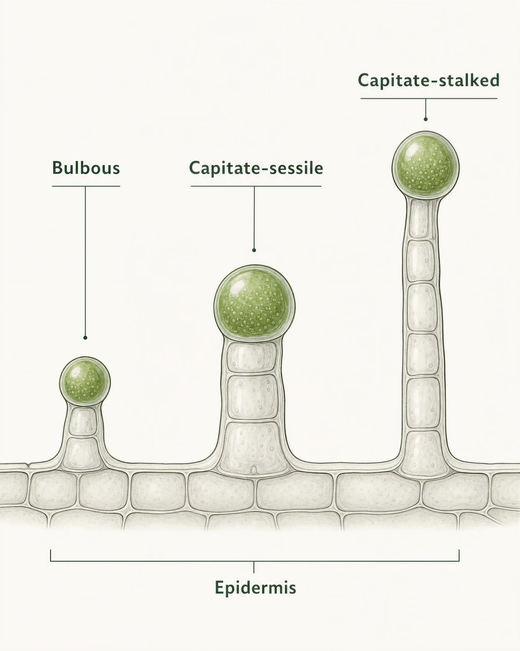
On cannabis, the three standard glandular trichome classes described in anatomical work by Hammond and Mahlberg and in later reviews are bulbous, capitate-sessile, and capitate-stalked. They are not interchangeable. They differ in size, architecture, and practical importance. On mature unfertilised female inflorescences, capitate-stalked trichomes are the major resin-producing form and the ones most tied to cannabinoid-rich floral material.
That point is not just botanical housekeeping. It changes how flower should be discussed. Paul Mahlberg and Eun S. Kim showed through microscopy that cannabinoids accumulate in the secretory cavity beneath the cuticle of glandular trichomes rather than being produced diffusely across all flower tissue. Happyana et al. in 2013 strengthened that localization argument using laser microdissection and mass spectrometry, showing cannabinoids and terpenoids concentrated in glandular trichomes. Livingston et al. in 2020 then added transcriptomic evidence: genes involved in cannabinoid biosynthesis are highly expressed in glandular trichomes of female flowers.
So trichomes are not decorative crystals sprinkled on a bud surface. They are tiny biochemical factories with storage compartments. Their morphology and integrity shape the chemistry people later measure, smell, and process.
Why trichomes matter to chemistry, harvest, and processing
Cannabis contains over 120 identified phytocannabinoids and more than 200 terpenes in the literature. The glandular trichome head is the primary site where much of that commercially and pharmacologically relevant chemistry is synthesized and stored. That alone explains why growers, processors, and scientifically literate consumers should care.
For growers, trichomes are a developmental indicator, but not a magic color code. Clear heads usually indicate immaturity. Cloudy or milky heads often align with a common harvest window associated with high THC accumulation. Amber heads point to later maturity and ongoing chemical change. Yet the popular rule that “amber means THC turned into CBN” is too tidy to be trusted as chemistry. Oxidation and degradation do occur, but color is a field signal, not a one-pigment, one-molecule equation.
For processors, trichomes matter even more directly. Kief, dry sift, bubble hash, and rosin are all trichome-centered products in one form or another. The condition of the gland head, the brittleness of the cuticle, the amount of contamination from non-gland tissue, and the maturity of the resin all affect what gets separated and what ends up altered.
Sinsemilla also makes more sense once trichomes are framed as reproductive-defense organs rather than glitter. Potter and Duncombe noted that unfertilised female floral bracts carry the highest densities of glandular trichomes. After pollination, plant resources shift toward seed production, and the intense resin-rich floral state becomes less pronounced.
Myths corrected in this article
- Myth 1 Visible frost does not equal potency; lab analysis settles cannabinoid strength.
- Myth 2 Not all trichomes are the same; bulbous, capitate-sessile, and capitate-stalked glands differ anatomically and functionally.
- Myth 3 Trichomes do not matter only at harvest; they matter during development, stress response, handling, and mechanical separation.
- Myth 4 More stress is not automatically better; UV-B and other stressors have conditional effects and can also harm output.
The common myths this article needs to correct
Visible frost equals potency. Limited evidence
The first myth is that visible frost equals potency. It does not. Dense trichome coverage can mean a flower is resinous, but potency is chemical, not optical. A cultivar with fewer visible trichomes can still produce higher cannabinoid concentration per gland head. Lab analysis, not surface sparkle, settles that question.
The second myth is that all trichomes are the same. They are not. Bulbous, capitate-sessile, and capitate-stalked trichomes differ anatomically and functionally, and reducing them to one generic “trichome” blurs real biology.
The third myth is that trichomes only matter at harvest. They matter throughout plant development, under environmental stress, during post-harvest handling, and in every mechanical separation method built around resin. Even the often-cited UV-B story needs restraint: Lydon, Teramura, and Coffman reported increased THC under enhanced UV-B in 1987, but that does not mean more stress always produces more resin or stronger flower.
This article treats trichomes as they should be treated: not as surface glitter, but as specialized glands that govern chemistry, maturity, and much of what people wrongly call quality at a glance.
| Trichome type | Typical size or profile | Structure | Usual distribution | Practical importance |
|---|---|---|---|---|
| Bulbous | Usually under about 20 micrometres in diameter | Small basal region with a very small glandular head | Broadly on aerial tissues | Not the main resin-bearing structure in harvested female inflorescences |
| Capitate-sessile | Larger than bulbous; low-profile gland | Basal cell, very short stalk region or compressed pedestal, multicellular glandular head | Leaves, bracts, and other surfaces | Biologically active, but usually less important than stalked glands on mature female flowers |
| Capitate-stalked | Large, conspicuous, mushroom-shaped resin gland | Basal cell, clear stalk, multicellular secretory head with subcuticular cavity | Concentrated on female floral organs, especially bracts | Main resin factory for mature unfertilised female flowers |

The three trichome types on cannabis
Cannabis does not produce one generic layer of “frost.” It produces three recognized glandular trichome types on aerial tissues: bulbous, capitate-sessile, and capitate-stalked. That classification comes from microscopy and histology work stretching from Paul G. Mahlberg and colleagues to later reviews and localization studies by Happyana et al. and Livingston et al. The distinction matters because these trichomes differ in size, cell arrangement, developmental timing, and resin output. If you flatten them into one category, you miss where cannabinoids and terpenes are actually concentrated.
Bulbous trichomes
Bulbous trichomes are the smallest glandular trichomes on cannabis. They are often described as tiny, almost microscopic protrusions that can be difficult to study without strong magnification. In practical terms, they are usually under about 20 micrometres in diameter, though measurements vary with method and tissue. They sit close to the epidermal surface and lack the dramatic mushroom-like profile associated with mature flower resin glands.
Anatomically, bulbous trichomes are simple. They consist of a small basal region anchored in the epidermis and a very small glandular head, often with only a few cells involved in secretion. Compared with the larger capitate forms, they have limited secretory volume. That means limited storage space beneath the cuticle and, as a result, far less resin accumulation visible to the eye.
Their practical importance is often overstated by articles that treat every sparkling point on the plant as equivalent. It is not. Bulbous trichomes may contribute to the plant’s protective chemistry, but they are not the dominant resin-bearing structures that define harvested female inflorescences. If the question is where the bulk of economically and horticulturally relevant cannabinoids are stored, bulbous trichomes are not the main answer.
Capitate-sessile trichomes
Capitate-sessile trichomes are larger than bulbous trichomes and clearly more developed as secretory organs. “Capitate” refers to the head, while “sessile” means they sit directly on the surface or on a very short stalk. Under magnification, they appear as rounded glandular heads attached close to the epidermis rather than elevated above it.
Subcuticular storage cavity The space that opens beneath the trichome cuticle as secreted resin accumulates. It acts as the main storage compartment of the gland head.
These trichomes have a more organized multicellular structure than bulbous glands. They include a basal cell, a short stalk region or compressed pedestal, and a glandular head made of secretory disc cells beneath a cuticular sheath. This is the architecture that begins to look like a proper resin gland rather than a minor epidermal outgrowth. As secretion accumulates, a subcuticular storage cavity forms between the secretory cells and the outer cuticle.
That storage pattern matters. Mahlberg and Kim’s microscopy showed that cannabinoids accumulate in the secretory cavity beneath the cuticle of glandular trichomes rather than being spread evenly across flower tissue. Happyana et al. reinforced the point in 2013 using laser microdissection and metabolite profiling, showing cannabinoids and terpenoids concentrated in glandular trichomes. Capitate-sessile glands participate in this secretory system, though they are usually less important than capitate-stalked glands on mature female flowers.
Developmentally, capitate-sessile trichomes tend to appear earlier and more broadly across plant surfaces than the large stalked glands associated with late flower maturation. They can be found on leaves and bracts, and they contribute to the chemical shield of the plant. Still, when growers or analysts care about resin-rich floral tissue, sessile glands are not the dominant feature.
Capitate-stalked trichomes
Capitate-stalked trichomes are the large, conspicuous resin glands most people actually mean when they talk about cannabis trichomes. These are the mushroom-shaped structures that become dense on mature, unfertilised female flowers. They have the most pronounced stalk, the largest glandular head, and the greatest secretory capacity of the three types.
Their anatomy is more elaborate. A basal cell anchors the structure in the epidermis. Above that sits the stalk, which elevates the glandular head away from the plant surface. At the top is the secretory head, made up of a multicellular disc that produces cannabinoids, terpenes, and other secondary metabolites. Those compounds are exported into the subcuticular cavity, where resin accumulates until the head appears swollen and glossy. Livingston et al. (2020) added transcriptomic support to the long-standing anatomical view by showing strong expression of cannabinoid biosynthetic genes in glandular trichomes, especially in flower tissues.
This is the trichome type with the greatest practical importance for harvested inflorescences. On mature female flowers, especially on the bracts surrounding the reproductive structures, capitate-stalked trichomes are the dominant resin-bearing glands. Potter and Duncombe’s cultivation and morphology work also pointed to unpollinated female inflorescences as the zone of highest glandular trichome density. That is the horticultural basis of sinsemilla production: keep flowers unfertilised, and the plant continues investing in resin-rich floral structures instead of shifting resources toward seed formation.
Where each type appears on the plant and why that distribution matters
The three trichome types are not distributed randomly. Bulbous trichomes occur broadly on aerial tissues, including stems and leaves, where they likely play a general protective role. Capitate-sessile trichomes also appear on vegetative tissues and smaller floral surfaces. Capitate-stalked trichomes, by contrast, become concentrated on female floral organs, especially bracts, during reproductive development.
That distribution is the reason harvested flower chemistry cannot be inferred from leaf frost alone. A sugar leaf may look glittery, yet the highest-value secretory structures are usually packed onto the floral bracts of mature female inflorescences. It also explains why male plants and non-floral tissues can have trichomes without producing the same resin load. Trichomes are not exclusive to female plants. Resin-rich capitate-stalked trichomes, densely concentrated on unfertilised female flowers, are what matter most in harvested material.
The hierarchy is clear. Bulbous trichomes are small and limited. Capitate-sessile trichomes are intermediate and biologically active. Capitate-stalked trichomes on mature female flowers are the main resin factories. That is the form most relevant to cannabinoid-rich inflorescences, and any serious discussion of trichomes has to start there.
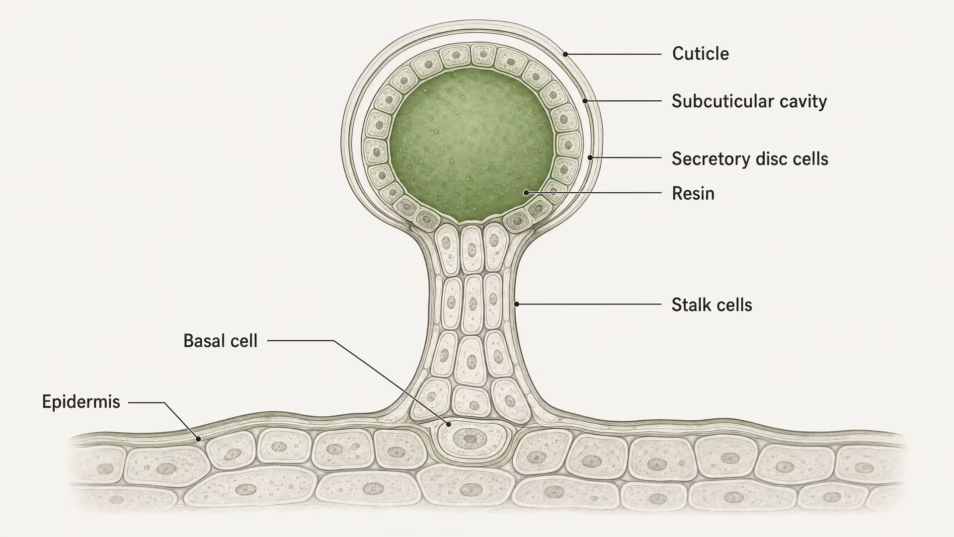
Anatomy of a resinous trichome
- Basal cell Anchors the gland in the epidermis and marks the point where epidermal tissue differentiates into a secretory appendage.
- Stalk Raises the glandular head above the surface in capitate-stalked trichomes and changes exposure and spacing.
- Secretory disc Contains metabolically active cells that synthesize cannabinoids, terpenes, and related metabolites.
- Cuticle-covered head Encloses the gland head and helps retain secreted material.
- Subcuticular cavity Stores resin beneath the cuticle until handling, rupture, oxidation, or processing alters the structure.

Trichome anatomy from basal cell to glandular head
When people talk about cannabis “frost,” they usually mean the swollen gland heads scattered across the floral surface. That shorthand misses the real biology. A resinous trichome is not a smear of oil coating the flower. It is a specialized epidermal organ with a defined architecture: an anchoring base, a stalk in some forms, a secretory disc of metabolically active cells, and a cuticle-covered head that stores secreted material in a distinct cavity. Histology and microscopy papers by Paul G. Mahlberg, Eun S. Kim, and later researchers made this clear decades ago. The gland head is where the action is.
Across cannabis, three glandular trichome classes are usually recognized: bulbous, capitate-sessile, and capitate-stalked. All share the same general logic of secretion, but capitate-stalked trichomes on mature female inflorescences are the dominant resin producers in practical terms. Their anatomy explains why.
The basal cell and epidermal anchoring point
At the bottom of the structure sits the basal cell, embedded in or arising from the epidermis. This is the trichome’s foundation. It anchors the entire gland to the outer tissue of the bract, sugar leaf, or other aerial surface and links the trichome physically and developmentally to the plant body.
The basal cell is not just a passive foot. In developmental terms, it marks the point where a regular epidermal cell lineage differentiates into a secretory appendage. As the trichome forms, this base establishes polarity: one end remains attached to the epidermal layer, while the upper region differentiates into stalk and head tissues. In capitate-stalked trichomes, that polarity is obvious under microscopy because the gland is lifted above the surface like a tiny mushroom. In sessile forms, the head appears much closer to the epidermis, but the same principle still applies.
Histological studies of cannabis trichomes show that these structures are organized rather than amorphous. Hammond and Mahlberg’s anatomical work, followed by Mahlberg and Kim’s ultrastructural studies, described the basal region as the point of insertion into epidermal tissue. This matters because the resin seen on a mature inflorescence does not originate as an exudate spread evenly through the flower. It emerges from discrete glandular units that are built from the epidermis upward.
The anchoring role of the basal cell also explains why trichomes can be mechanically detached. Kief, dry sift, and related separated resin fractions are composed largely of gland heads and associated fragments because the trichome is a mounted structure, not an internal reservoir diffused through plant tissue. Break the connection above the base, and the resin-bearing part can be removed.
The stalk and how it elevates the secretory head
The stalk is the most visually obvious difference between capitate-stalked and lower-profile gland types. In capitate-stalked trichomes, a column of stalk cells raises the glandular head above the epidermal surface. That elevation is not decorative. It changes exposure, spacing, and storage geometry.
On mature female flowers, the stalk acts like a pedestal for the secretory apparatus. By lifting the head away from the surface, the trichome can present a larger glandular sphere into the boundary layer around the flower. This likely improves the defensive value of the secretion. A raised, fragile gland is easier to rupture on contact by herbivores or handling, releasing sticky and chemically active contents where they are most likely to matter.
From an anatomical standpoint, the stalk consists of elongated cells situated between the basal cell and the head. In capitate-sessile trichomes, this segment is greatly reduced or nearly absent, which is why the gland appears seated directly on the epidermis. Bulbous trichomes are smaller still and far less important as resin reservoirs. Capitate-stalked trichomes, by contrast, combine height with a larger head and a larger secretory volume.
Microscopy work consistently shows that the biggest cannabinoid-rich glands on mature female inflorescences are these stalked forms. Potter and Duncombe’s cultivation morphology observations fit with this practical reality: unpollinated female floral bracts carry dense populations of the resinous glands that matter most for cannabinoid production. The stalk is part of that design. It spatially separates the biosynthetic and storage compartment from the living epidermal surface below, which may help both secretion and protection.
The secretory disc as the biochemical engine
Above the stalk sits the secretory disc, the cellular engine of the gland. This is the tissue that deserves far more attention than it usually gets. The disc is composed of secretory cells arranged beneath the outer cuticle, and these cells are metabolically specialized for the synthesis and export of the compounds later found in the resin cavity.
Cannabinoid biosynthesis is tightly associated with glandular trichomes, not with all flower tissues equally. Happyana et al. in 2013 used laser microdissection combined with mass spectrometry to show that cannabinoids and terpenoids are concentrated in glandular trichomes. Livingston et al. in 2020 strengthened this picture with transcriptomic evidence, showing high expression of cannabinoid biosynthetic genes in glandular trichome tissues of female flowers. That is why the gland head is not merely a storage bubble. It is a biosynthetic organ.
The secretory disc cells produce and export metabolites toward the space beneath the cuticle. In cannabis, this includes the pathway from olivetolic acid and geranyl pyrophosphate to cannabigerolic acid (CBGA), followed by enzyme-driven conversion into acidic cannabinoids such as THCA and CBDA in appropriate chemotypes. Terpene synthesis is also strongly represented in these glands. Reviews of cannabis chemistry now commonly cite more than 120 phytocannabinoids and over 200 terpenes identified across the species, and the glandular disc is central to where much of that specialized metabolism is organized.
This is the point popular writing often gets wrong. Resin is not simply “inside the flower.” It is made by secretory cells in localized epidermal glands. Trichome abundance can therefore matter, but only alongside gland size, developmental stage, and metabolic activity per gland.
The glandular head, subcuticular cavity, and resin storage
The glandular head is the swollen terminal structure most people inspect at harvest. Its defining feature is not color alone but architecture. Secretions produced by the disc cells accumulate beneath the cuticle, forming a subcuticular storage cavity. Mahlberg and Kim showed this clearly with microscopic and histochemical work: cannabinoids collect in this cavity under the distended cuticular sheath rather than dispersing uniformly across surrounding floral tissue.
That detail changes how trichomes should be understood. The visible “head” is a pressurized storage chamber capped by cuticle. As secreted resin accumulates, the cuticle separates from the underlying secretory cells, creating the cavity. The gland therefore has two related functions: biosynthesis in the disc cells and extracellular storage in the subcuticular space. The cuticle acts as the enclosing membrane-like barrier that retains the resin until mechanical rupture, senescence, oxidation, or processing alters the structure.
Under magnification, mature capitate-stalked trichomes often look like glassy globes, then cloudy spheres, and later darker or ambered heads. Those appearance changes are useful, but they are secondary signs. The primary fact is structural: if the head collapses, ruptures, oxidizes, or dries out, storage integrity is changing. That is often more biologically meaningful than a simplistic “amber equals better” rule.
The resin, then, is not spread as a uniform lacquer over the flower. It is compartmentalized inside thousands of microscopic gland heads, each mounted on its own epidermal base and, in the largest forms, elevated by a stalk. That arrangement explains almost everything that follows in cannabis handling and evaluation: why mature female bracts are resin-rich, why detached gland heads can be mechanically separated, why physical damage reduces quality, and why visible density alone does not prove chemical strength. The trichome head is both factory and vault.
Where cannabinoids and terpenes are made
Biosynthesis inside the glandular trichome head
Cannabinoids and most aroma-active terpenes are made primarily in glandular trichomes, especially the capitate-stalked trichomes that crowd mature female flowers. That statement is far more precise than the usual shorthand that “the plant makes THC in the buds.” The flower is the organ. The glandular trichome head is the main secretory factory.
Histology and microscopy work by Paul G. Mahlberg and Eun S. Kim helped establish the structural basis for this. In glandular trichomes, the head contains a disc of secretory cells covered by a cuticle. As metabolites are produced and exported, they accumulate in a subcuticular storage cavity. That matters because cannabinoids are not simply smeared through all floral tissue at equal levels. They are synthesized by specialized epidermal cells and stored outside those cells, under the lifted cuticle, in a resinous compartment.
The biosynthetic logic is plant-physiology 101, but with a cannabis-specific twist. Secretory disc cells are metabolically active, packed with plastids, vacuoles, smooth endoplasmic reticulum, and the enzymatic machinery needed for intensive secondary metabolism. These cells generate precursor molecules, run oxidocyclase reactions, and then move the products into the storage space. The glandular head is therefore both a synthesis site and a staging site for secretion.
This is why visible trichomes matter biologically. But they are not magical potency beads. A flower covered in resin can still test lower than a less frosty flower if the trichomes are genetically programmed to produce less THCA, CBDA, or terpene mass per gland head. Density and biosynthetic output are related only loosely.
The precursor pathway from CBGA to THCA and CBDA
The central precursor in major cannabinoid biosynthesis is cannabigerolic acid, or CBGA. It is formed when geranyl pyrophosphate, a terpenoid precursor, combines with olivetolic acid, a polyketide-derived precursor. This reaction links two metabolic streams: isoprenoid metabolism and fatty acid/polyketide metabolism. That hybrid origin is one reason cannabinoids do not fit neatly into a single classic plant-metabolite category.
Once CBGA is formed, cultivar genetics take over. The plant’s oxidocyclase enzymes convert CBGA into different acidic cannabinoids. THCA synthase produces tetrahydrocannabinolic acid. CBDA synthase produces cannabidiolic acid. A third route, via CBCA synthase, yields cannabichromenic acid. These acidic forms are the native plant products. Fresh cannabis does not biosynthesize large amounts of neutral THC or CBD directly. Those appear mainly after decarboxylation through heat, time, or processing.
This pathway has been worked out over decades, with chemistry reviews by ElSohly, Slade, and others clarifying the diversity of phytocannabinoids, while molecular studies identified the enzymes behind the major branches. What matters for trichome biology is location. These conversions are concentrated in the secretory tissues of glandular trichomes, not spread evenly across leaves, stems, and pistils.
There is also a practical implication. If one plant carries a highly active THCA synthase gene set, it can load CBGA toward THCA efficiently. Another genotype may favor CBDA. Another may do both poorly despite looking resinous. So a frosted appearance cannot, by itself, predict chemotype.
Terpene biosynthesis in the same secretory system
Terpenes are produced in the same general secretory architecture, which is one reason resin chemistry is such a tightly linked mixture rather than a pile of separate compounds. Cannabis contains more than 200 terpenes in the literature, though only a smaller subset usually dominates flower aroma. Monoterpenes such as myrcene, limonene, and pinene arise largely from the plastidial MEP pathway, while sesquiterpenes often draw from the cytosolic mevalonate pathway. In glandular trichome secretory cells, these pathways feed terpene synthase enzymes that generate the volatile profile.
Happyana et al. in 2013 provided some of the clearest direct evidence that terpenoids, alongside cannabinoids, are concentrated in glandular trichomes. Using laser microdissection and metabolite profiling, they showed that the trichome fractions carried the compounds most people associate with resin quality. This was not a visual observation. It was location-specific chemistry.
The shared secretory setting also helps explain why environmental conditions can change aroma and cannabinoid output together, though not always in parallel. A plant under altered light, temperature, or developmental conditions may shift the balance of both terpene and cannabinoid metabolism because both are being run by the same specialized cellular machinery.
What the localization studies actually proved
This is where the science often gets simplified too aggressively. The major localization studies did not prove that every cannabinoid in the plant exists only in one trichome type, nor did they prove that resin appearance is a direct potency meter. What they did prove is more useful.
Mahlberg and Kim’s anatomical studies showed that cannabinoids accumulate in the secretory cavity of glandular trichomes beneath the cuticle. That established the structural destination of the resin. Happyana et al. (2013) then used laser microdissection plus mass spectrometry to map phytocannabinoids and terpenoids to glandular trichome tissues with far greater specificity. Livingston et al. (2020), using transcriptomic and microscopic evidence, showed that cannabinoid biosynthetic genes are strongly expressed in glandular trichomes of female flowers. Put plainly: the trichome head is not just a storage bubble. It is a biosynthetic hotspot.
That still leaves room for nuance. “Hotspot” does not mean “independent of the rest of the plant.” The trichome depends on carbon supply, developmental signals, mineral nutrition, light environment, and genotype. If the plant lacks the genetic capacity to produce high THCA or high CBDA, no amount of visible frost changes that. The gland is a specialized output organ, not an isolated chemistry machine detached from breeding and physiology.
Cannabinoids and terpenes are made mainly in glandular trichome heads, especially on mature female inflorescences. Strong evidence
The strongest evidence, then, supports a balanced position. Cannabinoids and terpenes are made mainly in glandular trichome heads, especially on mature female inflorescences, by secretory cells that synthesize precursors, run enzyme-driven conversions, and export the products into a subcuticular cavity. That is the real biology behind resin. Not glitter. Not mythology. A specialized epidermal secretion system shaped by development and genetics.
Why sinsemilla flowers become resin-rich
Sinsemilla Seedless female cannabis flowers. In the article, the term is tied to unfertilised female inflorescences that keep investing in resin-rich glandular trichomes instead of shifting resources toward seed formation.
Sinsemilla means seedless female cannabis flowers, but the term only makes sense if you understand what the plant is doing biologically. Resin is not decorative frost. It is a secretion produced by glandular trichomes, especially the capitate-stalked glands concentrated on female floral bracts and the tissues immediately around them. When a female inflorescence remains unfertilised, it keeps investing in these glands. When pollination happens, that investment changes direction. The plant stops behaving like a flower still trying to attract pollen and starts behaving like a developing seed factory.
Unfertilised female inflorescences and reproductive strategy
An unfertilised female flower is still in reproductive limbo. It has produced stigmas to catch pollen, but until fertilisation occurs, the inflorescence remains metabolically active in ways that favor continued floral function and protection. That is the horticultural basis of the sinsemilla effect.
The highest resin densities in cannabis are found on unfertilised female bracts and adjacent floral tissues, not evenly across the whole plant. Potter and Duncombe, writing on cannabis cultivation morphology for the UK Home Office, described this concentration clearly: the bracts of seedless female inflorescences carry the richest covering of glandular trichomes. Those trichomes are not random outgrowths. They are specialized epidermal glands with secretory cells and a subcuticular storage space where cannabinoids and many terpenes accumulate.
Why would an unfertilised female keep making so much resin? Because the flower remains exposed and reproductively valuable. The bracts enclose ovules. The stigmas are still trying to intercept pollen. In that state, investing in glandular secretions likely serves several functions at once: defense against herbivores and pathogens, protection from UV stress, moderation of surface microclimate, and maintenance of a chemically active floral interface. Cannabis-specific work does not reduce resin to a single purpose, but the defensive interpretation fits the broader plant trichome literature well.
Modern localization studies support the idea that this investment is highly targeted. Happyana et al. (2013), using laser microdissection and metabolite profiling, showed cannabinoids and terpenoids concentrated in glandular trichomes. Livingston et al. (2020) added transcriptomic evidence showing strong expression of cannabinoid biosynthesis genes in glandular trichomes of female flowers. So the sinsemilla effect is not folklore. It reflects where the plant places secretory effort when reproduction is still unresolved.
What changes after pollination
Pollination changes the plant’s priorities fast. Once pollen lands, germinates, and fertilises the ovule, the female flower no longer needs to keep maximizing the same level of exposed floral signaling and glandular investment. Resources shift toward embryo and seed development.
That shift matters because plant metabolism is finite. Carbon skeletons, reducing power, mineral nutrients, and photosynthate cannot be spent twice. After pollination, more of that budget is routed into seed formation, bract swelling around developing seed, and maturation processes tied to reproduction rather than continued resin-heavy floral maintenance. Seeded flowers can still bear trichomes, but they generally do not keep building resin with the same intensity seen in unpollinated flowers.
This is why seedless cultivation became so important in resin-focused production. It is not that pollinated plants suddenly become trichome-free. They do not. The point is that fertilisation changes allocation. The plant has achieved reproductive success, so the selective pressure maintaining lavish glandular output on exposed female floral tissue is reduced.
A common oversimplification says pollination “stops THC production” outright. That is too blunt. What the evidence supports is a relative shift away from continued resin-rich floral development and toward seeds. In practical terms, that means less dense, less resinous inflorescences than comparable unpollinated female flowers.
Why male plants and leaves are different
Male plants can have trichomes. Leaves can too. But commercially significant resin density is concentrated elsewhere: on unfertilised female inflorescences, especially the bracts and nearby sugar leaves. That distinction matters because many popular explanations imply that only female plants make trichomes at all, which is false.
The difference is one of morphology, density, and function. Mature female flowers develop abundant capitate-stalked glandular trichomes, the form most associated with high cannabinoid and terpene accumulation. Male flowers generally produce fewer of these resin-rich glands, and their reproductive role is different. They are built to release pollen, not to maintain a long-lived, gland-heavy surface around unfertilised ovules. Leaves, meanwhile, often carry lower trichome densities and a different mix of gland types. They contribute far less to total resin output than female floral tissue.
Research by Mahlberg and Kim showed that cannabinoids accumulate in glandular trichome secretory cavities rather than diffusely across all tissues. That helps explain why “the plant” is not uniformly resinous. Resin production is anatomically localized, and the tissue most heavily endowed with the right glands is the unfertilised female flower.
So when growers talk about sinsemilla as resin-rich, the biologically accurate statement is narrower and sharper: seedless female inflorescences keep investing in glandular trichomes because reproduction is still pending, while pollination redirects development toward seeds. That is why the frosted look concentrates where it does, and why resin abundance is fundamentally a flower biology story, not a whole-plant one.
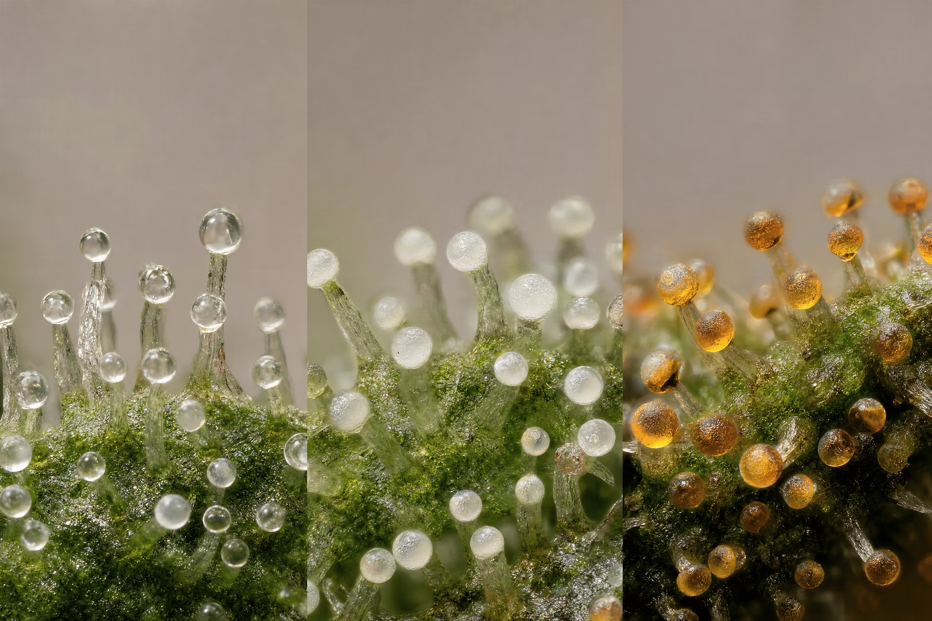
| Observed trichome state | What it usually signals | Common grower interpretation | Important caution |
|---|---|---|---|
| Clear | Immature gland heads and incomplete resin development | Wait | A frosty look does not mean the heads are mature |
| Cloudy or milky | Mature gland development and the usual peak harvest window | Go | Color alone cannot measure THC directly |
| Amber | Later maturity with oxidation and degradation processes | Late | Amber is not a simple one-color sign that THC became CBN |

Reading trichome maturity for harvest timing
Harvest timing advice often gets reduced to a color wheel: clear means wait, cloudy means go, amber means late. That shorthand is useful, but only if it is tied back to what trichomes actually are. These are glandular secretory structures, not glitter. In cannabis, the gland head of the capitate trichome is where cannabinoids and many terpenes are synthesized and stored, with classic anatomical work by Mahlberg and Kim and later localization studies such as Happyana et al. (2013) showing that cannabinoids are concentrated in glandular trichomes rather than evenly distributed across the flower.
That matters because “readiness” is not a mystical property of the bud as a whole. It reflects the developmental state of thousands of individual gland heads, especially on the bracts and calyxes of unfertilised female flowers, where resin-rich capitate-stalked trichomes are densest. If you want a practical framework, home growers are right to inspect trichome appearance directly. They are wrong when they turn that into a rigid myth, especially the claim that amber automatically means THC has turned into CBN. The real chemistry is messier.
Clear trichomes: immature glands and incomplete resin development
Clear trichomes usually indicate immature gland heads. Under magnification, the head appears glassy, transparent, and still “wet” looking rather than opaque. On a developing inflorescence, this stage generally corresponds to incomplete resin accumulation and a glandular head that has not yet reached full secretory maturity.
That does not mean no cannabinoids are present. It means the trichome is still in a developmental phase. Livingston et al. (2020) showed that cannabinoid biosynthetic activity is strongly associated with glandular trichomes, and trichome maturation is linked to changes in gene expression and secretory output. In practical terms, when most gland heads are still clear, the flower is usually still building toward its main cannabinoid peak rather than sitting at it.
Growers sometimes cut early because the plant already looks frosty. That is a visual trap. Resin abundance and resin maturity are not the same thing. A flower can be heavily covered in visible trichomes while many of those heads are still immature. This is one reason “frosty” appearance alone is a weak quality metric. Density does not tell you whether the secretory cavity is fully developed or whether cannabinoid concentration per gland head has peaked.
Inspecting clear trichomes also helps avoid another common mistake: judging by sugar leaves. Sugar leaf trichomes often mature earlier than the trichomes on the calyxes and bracts that make up the core of the flower. If sugar leaf heads are turning cloudy while bract trichomes remain mostly clear, harvesting at that point usually means cutting before the flower itself has finished developing.
Cloudy or milky trichomes: the usual peak harvest window
Cloudy or milky trichomes are the stage most growers target, and with good reason. The change from transparent to opaque reflects a mature gland head with dense resin content and altered light scattering through the secretory cavity. In practice, this phase often aligns with the period of highest THC potential.
“Often” is the word to keep. No microscope can directly measure THC concentration by eye, and no single trichome color guarantees a lab result. Still, the cloudy stage has become the standard harvest window because it usually corresponds to full gland development before later oxidative or degradative changes become more pronounced. This is not folklore pulled from nowhere; it fits the basic biology of gland maturation and resin accumulation described in cannabis anatomy and biosynthesis studies.
For home growers, the most reliable working rule is to inspect several parts of the plant, focusing on calyx or bract trichomes rather than leaf surfaces, and look for a majority of gland heads to be cloudy with only a minority still fully clear. A 30x loupe can show broad trends, but 60x to 100x magnification is much better for separating truly translucent heads from milky ones. At lower power, clear and cloudy can blur together.
This is also the point where expectations need to stay realistic. Cloudy trichomes do not mean every cultivar will produce the same effect profile. Cannabinoid ratios, terpene composition, and post-harvest handling all matter. ElSohly and Slade’s work on cannabis chemistry has long shown that cannabis is chemically varied, with far more going on than THC alone. So the cloudy stage is a practical harvest marker, not a universal promise.
Amber trichomes: late maturity, oxidation, and the CBN oversimplification
Amber trichomes are usually treated as the “late” end of the window, but popular explanations often get sloppy. The most repeated version says amber means THC has converted into CBN. That is too neat to be taken literally.
Amber coloration is better understood as a visible sign of later-stage chemical change in the gland head. Oxidation, degradation, and broader aging processes are involved. THC can degrade over time, and CBN is one known oxidation-related product, but fresh flower does not suddenly become CBN-rich just because a portion of the trichomes have turned amber. In most fresh material, CBN remains far below THC. The chemistry depends on cultivar, environment, handling, and time, not on a one-color-one-compound rule.
So why do growers watch for amber at all? Because it is still useful as a maturity marker. A small proportion of amber heads often suggests the plant has moved past peak immaturity and into a later harvest window. A large proportion generally signals that the flower is aging further, with greater risk of THC loss and terpene decline. That does not make amber “bad,” but it does make the simplistic “more amber equals stronger” claim unreliable.
The practical takeaway is moderation. If a grower wants the most common peak-THC-oriented window, mostly cloudy with limited amber is usually the target. If the flower is left much longer, the shift is better described as continued maturation plus degradation than as a clean THC-to-CBN switch.
Why pistil color is a weaker indicator than direct trichome inspection
Pistils are visible. Trichomes need magnification. That is why many growers still rely on pistil color first. The problem is that pistils are indirect. They can darken, curl, or recede for reasons that do not map cleanly onto gland maturity, including cultivar traits, environmental stress, pollination status, or simple handling.
A flower can show a high percentage of darkened pistils while many bract trichomes are still clear. The opposite can also happen. Pistils are part of the reproductive structure; trichomes are the secretory glands where the cannabinoids are actually made and stored. If the goal is harvest timing based on resin maturity, direct gland inspection is the stronger method every time.
Potter and Duncombe’s observations on inflorescence morphology are useful here because they reinforce where the most relevant glandular trichomes are concentrated: on the bracts of unpollinated female flowers. That is where the inspection should happen. Not just on the uppermost sugar leaves, and not by pistil color alone.
For home use, a simple routine works well: inspect multiple buds, sample middle and upper bracts, avoid judging from one flashy cola, and compare the ratio of clear, cloudy, and amber heads across the plant. That approach is imperfect, but it is far closer to the underlying biology than the old brown-hair harvest rule.
How environment shapes trichome density and resin output
UV-B exposure and the evidence behind the claim[1] UV-B radiation effects on photosynthesis, growth and cannabinoid production of two Cannabis sativa chemotypes. John Lydon, A. H. Teramura, C. B. Coffman. Photochemistry and Photobiology, 1987.
The idea that ultraviolet light makes cannabis “frostier” has a real scientific origin, but it has been stretched far past what the evidence actually shows. The paper almost always cited is Lydon, Teramura, and Coffman (1987), published in Photochemistry and Photobiology. In controlled conditions, they found that enhanced UV-B exposure increased delta-9-THC concentration in drug-type Cannabis sativa. That result matters. It suggests that UV-B can shift cannabinoid production under some conditions.
It does not prove a universal rule that stronger UV-B always means more trichomes, more resin, or stronger flowers.
That distinction matters because trichome density and trichome chemistry are separate variables. A plant can form many glandular heads yet produce less cannabinoid per gland than another genotype with fewer visible trichomes. Happyana et al. (2013) and Livingston et al. (2020) help frame the issue properly: cannabinoids and terpenoids are concentrated in glandular trichomes, and biosynthetic genes are strongly expressed in those structures, especially in mature female flowers. If UV-B changes resin output, it is likely doing so through stress signaling, altered secondary metabolism, or changes in gland development rather than by some simple “sunlight equals more crystals” mechanism.
There is also a broad plant-physiology rationale. UV-B is damaging radiation. Many plants respond by increasing protective surface compounds, pigments, or secretions that absorb, scatter, or reduce radiation damage. In cannabis, resin-rich glandular trichomes may contribute to that protective barrier. But genotype matters a lot. So do intensity, duration, developmental stage, leaf temperature, and overall plant health. Small increases in protective metabolite production are plausible. Excessive UV-B can just injure tissue, impair photosynthesis, and reduce flower development.
The restrained version is the accurate one: UV-B can alter cannabinoid accumulation in some experimental settings, but the effect is conditional, cultivar-dependent, and not a shortcut to quality.
Temperature swings, cold nights, and stress signaling
Grower folklore often treats cold nights as a resin trigger. The reality is less dramatic and more biologically believable. Temperature fluctuation can affect plant metabolism, membrane stability, enzyme activity, water relations, and stress hormone signaling. Those shifts can, in some genotypes, influence secondary metabolite production, including terpenes and cannabinoids. They can also affect trichome development indirectly by changing the pace of floral maturation.
That does not mean cold stress is automatically beneficial.
Cannabinoid biosynthesis depends on active cellular metabolism inside glandular trichomes. The secretory disc cells that feed the glandular head need functioning enzymes, energy supply, and intact membranes. Extreme cold can slow metabolism enough to reduce biosynthetic throughput. It can also increase the risk of tissue damage, stalled growth, and poor finishing. Temperature swings that are moderate may act as a mild abiotic signal. Swings that are severe tend to be counterproductive.
Research on cannabis environmental control still lags behind research on many major crops, so claims here should stay disciplined. Reviews by Andre, Hausman, Guerriero, and colleagues on cannabis morphology and specialized metabolism point toward environment-sensitive secondary metabolism, but they do not support the popular claim that sharply cold nights are a reliable resin hack. Sometimes cooler finishing temperatures help preserve volatile terpenes by reducing evaporative loss. That is not the same as saying they create more glandular resin.
One more point gets missed: temperature influences appearance. Cooler conditions can alter pigmentation and change the visual contrast between trichomes and floral tissue. A flower may look more dramatic without any meaningful rise in cannabinoid concentration. Visual frost is easy to overread.
Drought stress and defensive metabolite allocation
Water limitation is another area where a little plant science gets turned into bad advice. Mild drought stress can redirect plant resources toward defensive chemistry in some species. Cannabis is not exempt from that general rule. Under restricted water availability, plants often increase signaling molecules such as abscisic acid, alter carbon allocation, and shift growth away from expansion toward survival. In theory, and sometimes in practice, that can coincide with increased accumulation of certain secondary metabolites.
But drought stress is a trade-off, not a gift.
Glandular trichomes are metabolically expensive structures. Resin synthesis requires carbon skeletons, reducing power, and functioning secretory cells. If drought becomes strong enough to suppress photosynthesis, close stomata for long periods, and limit carbon assimilation, the plant has less raw material to build flowers and less energy to maintain resin production. You may see smaller yield, impaired floral development, and harsher post-harvest material even if a stress response has altered chemistry in some way.
This is where the distinction between concentration and total output becomes important. A stressed plant can sometimes show a higher concentration of a metabolite on a dry-weight basis while producing less total flower mass and less total cannabinoid overall. That is not the same thing as improved crop quality. It is often just stress concentrating what remains in reduced biomass.
Plant defense theory supports the idea that drought can change glandular behavior. Many aromatic plants increase protective or deterrent compounds under water stress. Yet the response is species-specific and genotype-specific, and timing matters. Early severe drought can permanently reduce plant capacity. Late mild deficit may shift chemistry without catastrophic yield loss. The simplistic idea that withholding water near the end automatically boosts resin is not supported as a general rule.
Evolutionary rationale: pest deterrence, UV screening, and microclimate buffering
Trichomes make the most sense when viewed as epidermal defense organs rather than sparkle. Across plant biology, glandular trichomes are associated with herbivore deterrence, pathogen defense, and protection from abiotic stress. Cannabis fits that broader pattern well. Resin is sticky, chemically active, aromatic, and positioned on exposed reproductive surfaces. That is exactly where a plant would place a defensive secretion.
Pest deterrence is the most intuitive role. A glandular head can physically impede small herbivores and deliver deterrent compounds at the tissue surface. Terpenes and cannabinoids are not there for human appreciation. They are part of a protective chemical interface. Reviews on glandular trichomes in aromatic and medicinal plants repeatedly support this defensive framing, and cannabis-specific work has long pointed in the same direction.
UV screening is also plausible. The Lydon study gave this idea its cannabis-specific foothold, but the broader concept comes from plant stress physiology: exposed reproductive tissues benefit from surface compounds that reduce radiation damage. Resin may absorb or scatter part of that burden.
Microclimate buffering is less discussed but biologically sensible. Dense trichome layers can alter the immediate boundary layer at the plant surface, affecting heat exchange, moisture loss, and tissue exposure. They are not miniature insulation blankets in a simplistic sense, yet they can modify the physical environment right where the plant is most vulnerable. On female inflorescences, where reproductive success matters, such buffering may have adaptive value.
This defensive framing also helps explain why unfertilised female flowers become especially resin-rich. As Potter and Duncombe noted, unpollinated female inflorescences carry the highest densities of glandular trichomes on the floral bracts. Once pollination occurs, resource allocation shifts toward seed production. Resin-heavy floral investment becomes less pronounced because the reproductive task has changed.
Why more stress is not always better
The popular mistake is thinking of stress as a knob you can keep turning upward. Biology does not work that way. Mild stress can induce protective responses. Strong stress can overwhelm them.
That is the key correction.
UV-B can increase THC under some controlled conditions. Cold or fluctuating temperatures can shift metabolism in some genotypes. Water deficit can alter defensive compound allocation. None of those findings justify the blanket claim that harsher conditions produce better flowers. At some point, stress reduces photosynthesis, damages membranes, stalls development, lowers yield, increases susceptibility to disease, and degrades the integrity of glandular heads themselves.
Resin output is the product of genetics, developmental stage, and environment working together. Environment can modulate the system. It does not override it. A cultivar with poor cannabinoid biosynthetic capacity will not become chemically exceptional just because it was stressed. Livingston et al. (2020) showed how strongly cannabinoid production is tied to glandular trichome biology and gene expression. That biology has limits.
The practical takeaway is plain: controlled, moderate environmental signaling may influence trichome density or metabolite composition, but stress beyond the plant’s coping range usually lowers overall quality. Frostier-looking material is not automatically more potent, and a harsher grow is not automatically a smarter one.

Microscopy for home growers: how to inspect trichomes properly
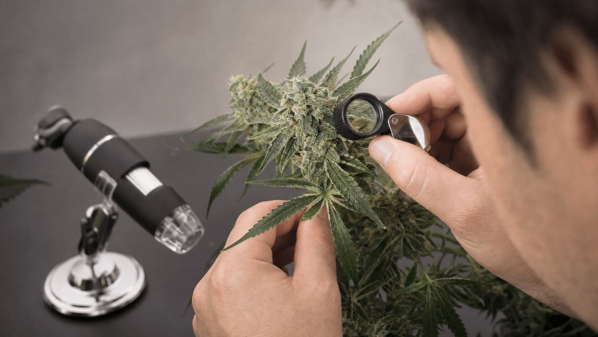
Trichome inspection is often reduced to a color check. That is too simple. You are looking at secretory glands, not glitter, and your goal is to judge gland-head development on the flower tissue that matters most. The practical target is the capitate-stalked glandular head on the bracts of mature female flowers, because that is where cannabinoids and terpenes are concentrated, as shown by Mahlberg and Kim’s anatomical work and later localization studies such as Happyana et al. (2013) and Livingston et al. (2020).
| Tool | Typical use in the article | Strengths | Limits |
|---|---|---|---|
| Jeweller's loupe | Broad maturity calls at about 30x | Cheap, portable, fast | Weak stability; glare and movement can blur clear versus cloudy heads |
| Digital microscope | Image capture and repeat comparisons at about 60x to 100x | Creates records and supports side-by-side checks | Awkward positioning in a live canopy without support |
| Clip-on phone optics or dedicated trichome scope | Quick close inspection with built-in lighting or phone capture | Convenient for repeat checks | Optical quality varies; reflections and blur can mislead |
Jeweller's loupe: cheap, portable, good enough for broad maturity calls
A basic loupe is still useful. At 30x, you can usually tell whether trichomes are mostly clear, mostly cloudy, or entering a later mixed stage. That is enough for broad harvest timing. It is not enough for fine calls on individual heads.
The strengths are obvious: low cost, no batteries, pocketable, fast. The weakness is stability. If your hand shakes, the flower moves, and the loupe has weak lighting, clear heads can look cloudy and glare can look amber. Many growers blame the tool when the real problem is motion.
Use the loupe on a still branch, ideally with the plant supported and airflow off. Inspect several sites, not one cola tip. Focus on the swollen bracts, not sugar-leaf tips. Leaf trichomes often amber earlier, get damaged more easily, and can push you into harvesting too soon.
Digital microscopes: better records, harder ergonomics
Digital microscopes are better when you want documentation. You can capture images, compare changes over several days, and avoid the “I think it looked milkier yesterday” problem. That makes them useful for side-by-side checks across cultivars or different canopy levels.
They are not automatically easier. In a live canopy, many USB and handheld digital scopes are awkward to position. The device, the cable, your hand, and the branch all want to move at once. Without a stand or a way to brace the scope, image quality drops fast. Good records require good support.
A digital microscope in the 60x to 100x range is usually enough for home inspection. Past that, magnification sounds impressive but often becomes less practical because field of view shrinks and shake becomes severe.
Dedicated trichome scopes and clip-on optics
Dedicated trichome scopes sit between a loupe and a digital microscope. They are designed for close inspection, often with built-in LEDs and fixed magnification. For many home growers, they are the easiest way to get repeatable looks at live flowers without juggling a phone and separate lens.
Clip-on phone optics can work, but quality varies a lot. Cheap lenses often add edge blur, color fringing, and reflections that make resin heads look stranger than they are. If you use one, clean the lens first and test it on known material before trusting your harvest call.
Magnification ranges and what to look for in live flowers
At 30x, expect trend reading. You can see whether heads are broadly transparent or broadly opaque. At 60x, the distinction between clear and cloudy becomes more reliable, and you can spot collapsed or broken heads. At 100x, you can inspect head shape, stalk attachment, and whether apparent amber is true pigmentation or just warm light, oxidation on damaged resin, or surface contamination.
Lighting matters as much as magnification. Cool, diffuse light is easier to read than a harsh point LED blasting straight into the resin. Change the angle slightly. If the “amber” disappears when the glare shifts, it was glare. If a head looks brown, check whether it is ruptured or dust-coated before calling it mature.
Look for patterns, not outliers. Sample upper, middle, and lower flowers. Prioritize intact gland heads on bracts. Ignore a few damaged trichomes unless they are representative of the whole flower. And remember the big limitation: microscopy can show maturity state and gland condition, but it cannot tell you potency. Frostier-looking flowers are not automatically stronger. Only chemical testing can answer that.
| Product or method | Core process | What happens to gland heads | Main quality variables |
|---|---|---|---|
| Kief / dry sift | Mechanical screening of dried material | Many heads detach and remain partly intact if handled well | Starting material maturity, screen cleanliness, plant contamination |
| Bubble hash | Ice-water agitation and filtration through mesh bags | Heads detach in cold water; some breakage can occur during agitation and drying | Agitation intensity, starting material type, filtration, drying |
| Rosin | Heat and pressure applied to flower, sift, or hash | Glands are collapsed to express a resinous phase | Input cleanliness, press temperature, pressure, terpene loss risk |
Trichome-derived products: from detached glands to pressed resin
Trichome products begin with a simple biological fact: cannabinoids and many terpenes are concentrated in glandular trichome heads, especially the capitate-stalked glands that dominate mature unfertilised female flowers. Mahlberg and Kim’s microscopy work, followed by direct localization studies such as Happyana et al. (2013), showed that these compounds are associated with the secretory structures of the gland rather than spread evenly through all floral tissue. Processing methods are therefore attempts to isolate, preserve, or rupture those glands in controlled ways. The differences between kief, hash, and rosin are mostly differences in how trichomes are separated and what happens to the gland head afterward.
Kief and dry sift
Kief is the loose granular material obtained when brittle trichome heads detach from dried flower and pass through a screen. Dry sift is the more deliberate version of the same idea: dried plant material is agitated across one or more mesh sizes so detached glands fall through while larger pieces of leaf and floral tissue are held back. This is mechanical separation, not extraction in the chemical sense.
The starting material matters. Well-dried flower or trim with mature, intact gland heads will release more usable resin than underdeveloped material with many clear trichomes or overhandled material where heads have already ruptured and smeared onto plant surfaces. Maturity affects both chemistry and behavior during screening. Cloudy heads tend to be fuller and less watery in appearance than clear immature heads, while heavily oxidized or degraded glands may break apart too easily and contaminate the sift with non-glandular debris.
Quality in dry sift is tied to cleanliness as much as yield. A pile of pale, sandy trichome heads and stalk fragments is not the same thing as green, dusty material full of pulverized leaf. Visible frost on the original flower can mislead here. Dense trichome coverage may produce a large volume of sift, but if those glands contain less cannabinoid per head, or if the sift includes substantial plant contamination, appearance outpaces chemistry. Trichome density and potency are related only loosely.
Bubble hash and ice-water separation
Bubble hash also starts with detached trichomes, but the route is different. Instead of dry screening, the material is stirred or agitated in very cold water with ice, then filtered through progressively finer mesh bags. The cold makes trichomes more brittle and less sticky, helping gland heads snap off from the epidermal surface. Water itself does not dissolve cannabinoids efficiently, so the process is still considered solventless in common use, though it is better described as ice-water mechanical separation.
Fresh-frozen and dried material behave differently. Fresh-frozen flower can retain a broader volatile profile because it avoids a full drying phase before separation, but it is also more technically demanding. Dried starting material is easier to handle, though terpene loss may already have occurred before washing begins. In both cases, the target is the same: separate intact or near-intact gland heads while limiting contamination from broken leaf tissue, pistils, cuticle fragments, and oxidized resin.
Agitation is a balancing act. Too little leaves resin behind. Too much shreds plant material and lowers purity. This is where trichome anatomy matters in practical terms: the gland head is a cuticle-enclosed secretory structure, and once that structure breaks, its contents can smear, oxidize, and trap debris. Bubble hash quality therefore reflects not just cultivar and harvest stage, but how gently the glands were detached and how well they were filtered afterward.
Rosin from flower, sift, or hash
Rosin is produced by applying heat and pressure to resin-bearing material so oily constituents flow out of the compressed mass. Unlike kief or bubble hash, which are primarily separation methods, rosin is an expression method. It does not detach intact glands for collection; it collapses them.
The starting material can be flower, dry sift, or hash. Flower rosin begins with resinous inflorescences and usually carries more waxes and plant compounds because the glands are being pressed while still embedded in flower tissue. Sift rosin starts from mechanically separated trichomes, while hash rosin starts from ice-water hash that has already undergone one purification step. That difference explains why the cleanliness of the input has such a strong effect on the output. Cleaner glands in, cleaner resin out.
Heat is both useful and destructive. It lowers viscosity and helps resin flow, but it also accelerates terpene evaporation and chemical change. Press too cool and yield may be poor. Press too hot and aromatic compounds disappear faster, while darker color and a more cooked profile become more likely. Rosin is still a trichome product, but it is no longer an intact-gland product.
What processing does to gland integrity and terpene retention
Every processing route trades something away. Kief and careful dry sift can preserve much of the physical identity of detached trichome heads, especially when the material is cold, dry, and handled lightly. Bubble hash can isolate glands effectively, but agitation and water movement can break fragile heads, and subsequent drying is another point where terpene loss or oxidation can occur. Rosin preserves the solventless principle yet intentionally destroys gland structure to express a resinous phase.
Handling quality often matters more than people admit. Warm fingers, repeated shaking, rough trimming, and poor storage all rupture gland heads before any intentional processing starts. Once the cuticle is broken, terpenes volatilize more readily and the sticky resin captures contaminants. That is why mature but not over-oxidized starting material usually performs better than either immature flower full of clear heads or old material with many collapsed amber glands.
A final correction is needed here. Product quality is not predicted by visible frost alone. It depends on trichome maturity, gland chemistry, physical integrity, contamination level, and post-harvest handling. The gland is the unit that matters. Processing either separates it, filters it, or crushes it.
Why trichome density is not the same thing as potency
A frosty flower can be impressive under light, but appearance is not chemistry. That distinction matters. Trichomes are secretory glands, not glitter, and potency is a chemical measurement of cannabinoids in the finished material, not a visual score based on how white or “sugary” the surface looks. The popular shortcut — more visible trichomes equals stronger flower — fails often enough that it should be treated as a myth, not a rule.
Peer-reviewed work on cannabis anatomy helps explain why. Mahlberg and Kim showed that cannabinoids accumulate in the secretory cavity of glandular trichomes, beneath the cuticle, rather than uniformly across all floral tissue. Happyana et al. (2013) later used laser microdissection and metabolite profiling to show that cannabinoids and terpenoids are concentrated in glandular trichomes. Livingston et al. (2020) added transcriptomic evidence showing strong expression of cannabinoid biosynthetic genes in trichome-rich female flower tissues. Those findings support a simple point: what matters is not only how many glands you can see, but what each gland has produced, stored, and retained.
Visual density versus cannabinoid concentration per gland
Two flowers can look very different and still reverse expectations in lab testing. One may carry a thick visible layer of trichomes yet produce only moderate total THC or CBD. Another may appear less dramatic but test higher because its gland heads are larger, more chemically productive, or more densely packed on the highest-value tissues such as the bracts rather than the sugar leaves.
Visible density is a blunt tool for several reasons. First, trichome heads vary in size and development. A flower covered in small, immature, clear gland heads may look heavily frosted but still be biochemically behind a less flashy flower with mature, cloudy capitate-stalked trichomes. Second, “frost” includes visual input from plant tissue. White pistils, reflective cuticle, and dense sugar leaf coverage can exaggerate the impression of resin abundance. Third, potency is measured against the weight of the harvested material. More leaf mixed into the sample can dilute cannabinoid percentage even if the surface looks resinous.
This is where the common confusion starts: resin abundance and cannabinoid concentration are related, but they are not identical. A cultivar can produce many glands whose contents are relatively moderate in THC. Another can produce fewer visible glands with higher cannabinoid concentration per gland head. ElSohly and Slade’s chemistry work has long underscored how complex cannabis composition is; over 120 cannabinoids and more than 200 terpenes have been identified in the literature. Trichomes are chemical factories, and factories differ in output.
Genetics, maturity, and post-harvest handling as hidden variables
Genetics sets the ceiling. Some cultivars are simply predisposed to produce more THC, more CBD, a different terpene profile, or different trichome morphology. Potter and Duncombe’s cultivation work, along with later anatomical reviews, showed that unfertilised female inflorescences carry the heaviest densities of the resin-rich glandular trichomes that matter most for cannabinoid production. Even within that category, cultivar differences are large. A dramatic-looking flower from one genotype may test below a less showy flower from another.
Maturity also changes the equation. Clear trichomes usually indicate immature glands. Cloudy or milky heads generally mark the main harvest window associated with peak THC accumulation. Amber heads suggest later maturity and chemical change, but the popular claim that amber simply means THC has turned into CBN is too neat to be fully accurate. Degradation and oxidation are real; the one-color, one-molecule story is not. A flower that looks extra “dusty” because many heads are aging, collapsing, or oxidising is not necessarily gaining potency.
Post-harvest handling may be the most overlooked variable of all. Heat, oxygen, light, and rough handling can damage gland heads and alter their contents after harvest. THC can degrade over time, terpenes can volatilise, and brittle trichome heads can break off. So a sample that once looked and tested strong may lose potency if drying, curing, or storage is poor. Visual frost tells you little about what has already degraded.
Why lab testing beats visual guesswork
Potency is a laboratory question. It is best answered by validated chemical analysis such as HPLC, which quantifies cannabinoids directly rather than inferring them from appearance. That is not pedantry. It is the only reliable way to separate dense-looking resin coverage from actual cannabinoid percentage.
Visual inspection still has value. It can help assess maturity, gland integrity, contamination, and rough handling damage. Under magnification, a grower can distinguish clear heads from cloudy ones and spot oxidised or ruptured trichomes. What visual inspection cannot do is calculate cannabinoid concentration with confidence. No loupe can tell you whether one cultivar’s gland heads contain substantially more THC or CBD than another’s.
The editorial position here should be firm: frosty appearance is an imperfect proxy, not a potency test. Trichome density can suggest careful cultivation, strong resin production, or good harvest timing, but it cannot settle potency on its own. Chemistry does that. When the question is strength, lab data beats guesswork every time.
What trichome science still does not answer cleanly
Limits of current cannabis trichome research
Cannabis trichome science is stronger than internet folklore suggests, but it is still thinner than many readers assume. We do have solid anatomy and localization work. Mahlberg and Kim showed that cannabinoids accumulate in the subcuticular secretory cavity of glandular trichomes rather than diffusely across all floral tissue. Happyana et al. (2013) then used laser microdissection and metabolite profiling to show cannabinoids and terpenoids concentrated in glandular trichomes. Livingston et al. (2020) added transcriptomic evidence that cannabinoid biosynthetic genes are highly active in these glands. That is a strong mechanistic base.
What remains messy is prediction. Research often uses specific cultivars, controlled environments, and narrow endpoints. A finding that applies to one genotype under one light spectrum may not scale cleanly to another. Lydon, Teramura, and Coffman’s 1987 UV-B paper is the classic example: it supports the idea that UV-B can alter THC production under some conditions, not the stronger claim that extra UV-B always increases resin, potency, or flower quality. The same caution applies to drought stress, temperature swings, and late-flower stress. Plants respond. Not always in the same direction, and not always beneficially.
Another limit is that visible trichome assessment still outruns chemical measurement in popular discussion. A trichome head can look abundant yet carry different cannabinoid and terpene profiles depending on genetics, maturity, and handling. Frost is morphology. Potency is chemistry.
Where grower heuristics are useful but not precise
Grower heuristics survive because many of them are directionally right. Clear trichomes usually indicate immature glands. Cloudy or milky heads often align with a common harvest window. More amber usually signals later maturity and chemical change. Unpollinated female flowers do tend to remain the major site of dense capitate-stalked resin production, which fits the sinsemilla principle described by Potter and Duncombe. These rules are practical.
Still, they are easy to overstate. “Amber means THC turned into CBN” is too neat. Oxidation and degradation do occur, but fresh flower does not suddenly become CBN-rich because some heads changed color. “More stress means more trichomes” is also too blunt. Moderate stress can increase defensive secondary metabolism in some cases; excessive stress can reduce yield, damage tissue, and lower total resin output. Even the old “more sparkle equals stronger flower” claim fails basic chemistry. Dense gland coverage may look impressive while biosynthetic output per gland remains modest.
Home microscopy has the same limitation. A 30x loupe can show broad trends. A 60x to 100x scope is better for distinguishing translucent, opaque, collapsed, or oxidized heads. Neither can replace cannabinoid analysis.
The strongest evidence-based takeaways
The firmest takeaway is structural: cannabis trichomes are specialized epidermal secretory organs, not cosmetic frost. Their class, anatomy, and developmental state matter. Bulbous, capitate-sessile, and capitate-stalked glands are not interchangeable, and the capitate-stalked form on mature female inflorescences does most of the heavy lifting for cannabinoid-rich resin.
The next firm point is chemical: localization matters more than sparkle. Cannabinoids and many terpenes are made and stored in glandular tissues, especially the head. That means harvest judgment should consider gland maturity and integrity, not color alone.
Beyond that, honest uncertainty is the right ending. Science supports some grower instincts, but often in softer terms than the culture prefers. Trichomes reward close looking, yet they resist simple rules. Anatomy, chemistry, genotype, and environment all shape what those tiny glands are doing. Sometimes a cloudy head means “ready.” Sometimes it only means “cloudy.”
References
- [1] UV-B radiation effects on photosynthesis, growth and cannabinoid production of two Cannabis sativa chemotypes. Photochemistry and Photobiology, 1987. https://doi.org/10.1111/j.1751-1097.1987.tb04757.x