













What the endocannabinoid system actually is
Not a cannabis receptor: correcting the most common misconception
The first correction matters because so many cannabis explainers get it wrong: the endocannabinoid system did not evolve for cannabis. It is not a receptor lock waiting for THC to arrive and turn it on.
What the ECS is actually built from
Core parts of the ECS
- Ligands Endocannabinoids such as anandamide (AEA) and 2-AG.
- Receptors Mainly CB1 and CB2, plus related non-cannabinoid targets in the wider network.
- Synthetic machinery Enzymes that make endocannabinoids on demand from membrane lipids.
- Transport and confinement Local mechanisms that keep signalling short-range.
- Catabolic enzymes FAAH, MAGL, ABHD6, and ABHD12 terminate signalling.
The ECS is an endogenous signalling network built from lipid messengers, their receptors, the enzymes that make them, the mechanisms that move and confine them, and the enzymes that shut them down.
How cannabis entered the ECS story — and why that changes the frame
Cannabis entered the story because plant cannabinoids happened to interact with that network strongly enough for scientists to notice. The system itself was already there, regulating local physiology across the brain, immune tissues, gut, endocrine organs, and more.
How the ECS was discovered through cannabis research
The endocannabinoid system was not discovered because scientists went looking for a built-in “cannabis pathway.” It emerged the way many hidden signalling systems do in pharmacology: a plant compound produced reproducible effects, researchers inferred that a specific molecular target must exist, then the body’s own ligands were found later. That sequence matters. The ECS is an endogenous lipid signalling network that cannabis happens to perturb. Historically, cannabis was the probe that exposed it.
From THC isolation to receptor hunting
The modern story starts with chemistry, not receptors. In 1964, Raphael Mechoulam and Yechiel Gaoni reported the isolation and structural elucidation of delta-9-tetrahydrocannabinol, or THC, from Cannabis sativa. Earlier investigators had identified cannabinoids such as cannabidiol, but THC was the major psychoactive constituent that could finally be studied as a defined molecule rather than as part of a crude plant extract. That changed the field.
Once THC could be purified and administered in controlled settings, a basic question became unavoidable: how was it producing its effects? By the 1970s and early 1980s, researchers knew THC altered memory, motor control, pain processing, appetite, and mood. Those effects were too selective, too anatomically patterned, to be explained well by a vague idea of membrane disruption. Lipid-soluble drugs can affect membranes, yes, but that could not account for the stereoselectivity seen with cannabinoids. Some cannabinoid analogues were much more potent than others, and tiny changes in molecular structure changed biological activity in predictable ways. That is classic receptor pharmacology.
Allyn Howlett’s work was especially important here. In the 1980s, her lab produced binding and signalling evidence that cannabinoids acted through a specific receptor coupled to G proteins. In 1988, Devane, Dysarz, Johnson, Melvin, and Howlett reported in Molecular Pharmacology the identification of a cannabinoid receptor in rat brain using the synthetic cannabinoid radioligand [3H]CP-55,940. This was the bridge between behavioral pharmacology and molecular biology. THC was no longer just a psychoactive plant compound. It had a high-affinity binding site in the brain.
That finding pushed the field into receptor hunting. If a receptor existed, where was it expressed? What kind of receptor was it? And, most important, why would the brain contain a receptor for a plant chemical at all? That last question was the giveaway. Biology does not evolve a receptor so humans can respond to cannabis. The obvious implication was that the receptor’s real ligands were endogenous and still unknown.
This is a recurring pattern in drug discovery. Opioid receptors were identified before endorphins. Benzodiazepine binding sites were characterized before endogenous modulators were sorted out. The cannabinoid field followed the same logic. Receptor first, native ligand second.
CB1 in 1990 and CB2 in 1993[1] Structure of a cannabinoid receptor and functional expression of the cloned cDNA. Lisa A. Matsuda, Stephen J. Lolait, M. J. Brownstein, A. C. Young, Thomas I. Bonner. Nature, 1990.
G protein-coupled receptor (GPCR) A cell-surface receptor that signals through intracellular G proteins. CB1 is a GPCR linked mainly to Gi/o proteins.
The first major breakthrough came in 1990, when Lisa Matsuda and colleagues cloned the cannabinoid receptor now called CB1, publishing the work in Nature. That paper established CB1 as a seven-transmembrane G protein-coupled receptor, primarily linked to Gi/o proteins. Functionally, that meant cannabinoid signalling could inhibit adenylyl cyclase, regulate ion channels, and suppress neurotransmitter release. Mechanistically, the field had moved from “THC does something to the brain” to a defined receptor and signalling architecture.
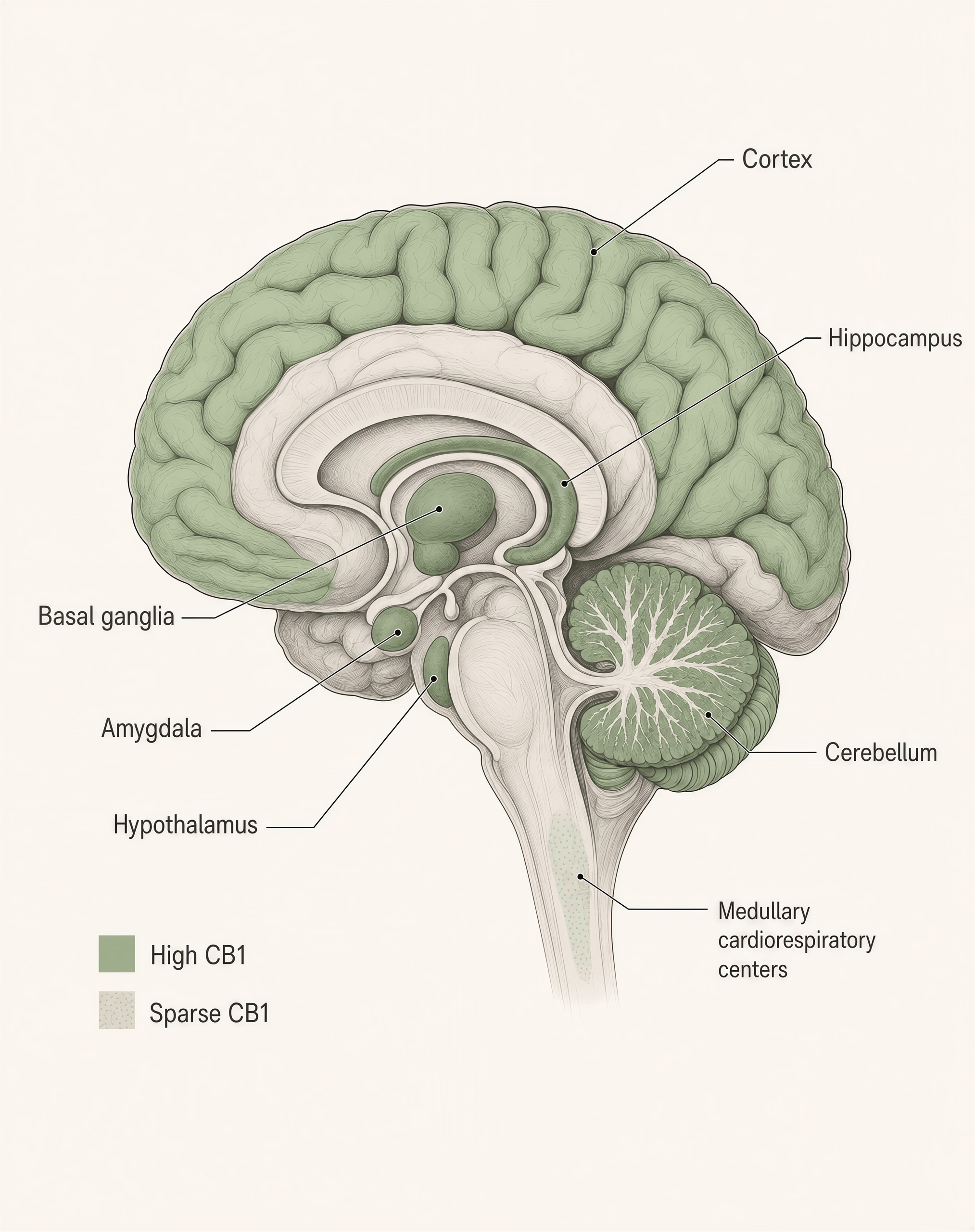
CB1’s expression pattern immediately helped explain major features of cannabis intoxication. It is highly expressed in cortex, hippocampus, basal ganglia, cerebellum, and limbic circuitry—regions tied to memory, timing, reward, movement, and affect. It is also one of the most abundant GPCRs in the brain. At the same time, CB1 expression is comparatively sparse in the medullary cardiorespiratory centers of the brainstem. That distribution is one reason cannabinoid overdose does not typically produce the fatal respiratory depression seen with opioids. The receptor map matched the pharmacology.[2] Molecular characterization of a peripheral receptor for cannabinoids. Sean Munro, Karen L. Thomas, Mona Abu-Shaar. Nature, 1993.
Then came CB2. In 1993, Sean Munro, Karen Thomas, and Mona Abu-Shaar cloned a second cannabinoid receptor, published in Nature. CB2 showed a very different expression profile from CB1, with prominent expression in immune cells and immune-related tissues rather than broad neuronal abundance. That finding reshaped the entire field. Cannabinoid biology was not only about psychoactivity. It had immunological dimensions too.
| Feature | CB1 | CB2 |
|---|---|---|
| Cloned | 1990 | 1993 |
| Main baseline pattern | Highly expressed in brain circuits | Enriched in immune cells and lymphoid tissues |
| Common shorthand | Brain receptor | Body receptor |
| Article's correction | Also present in peripheral tissues | Can become relevant in CNS inflammation |
Meaningful neuronal CB2 expression in the healthy brain remains debated and context-dependent. Limited evidence
Popular summaries often freeze the story there: CB1 equals brain, CB2 equals body. That is too tidy. CB1 is indeed dominant in central synaptic signalling, but it is also found in peripheral tissues. CB2 is enriched in immune compartments, yet low-level expression in parts of the nervous system can appear under inflammatory or pathological conditions, and the extent of true neuronal CB2 expression remains context-dependent and still debated. Even in the 1990s, the lesson was already that cannabinoid signalling involved distribution patterns, not cartoon categories.
The cloning of CB1 and CB2 also sharpened the central mystery. If mammals express not one but two cannabinoid receptors, then THC is almost certainly imitating a preexisting signalling language. Researchers now had the receptors. The next step was to find the endogenous words.
The discovery of anandamide and 2-AG[3] Isolation and structure of a brain constituent that binds to the cannabinoid receptor. William A. Devane, Lumír Hanuš, Aviva Breuer, Raphael Mechoulam. Science, 1992.
That search succeeded quickly. In 1992, William Devane, Lumír Hanuš, Allyn Howlett, Raphael Mechoulam, and colleagues identified the first endogenous cannabinoid ligand, arachidonoylethanolamide, better known as anandamide or AEA, in Science. The name came from the Sanskrit ananda, meaning bliss, paired with the chemical suffix for amide. The name made headlines, but the chemistry was the real turning point.
Anandamide was not stored in synaptic vesicles like a classical neurotransmitter. It was a lipid-derived signalling molecule made from membrane precursors. It was also short-lived. That hinted from the start that the ECS would not look like dopamine or serotonin systems. It would be more local, more transient, and more tightly tied to membrane lipid metabolism. AEA bound CB1 and helped explain why the receptor existed at all: the brain had its own cannabinoid-like messenger.
Yet anandamide was only part of the picture. In many tissues, especially brain, it turned out not to be the quantitatively dominant endocannabinoid. In 1995, two groups independently advanced the next major step. Mechoulam and colleagues identified 2-arachidonoylglycerol, or 2-AG, as an endogenous cannabinoid ligand, while Tomoyuki Sugiura and colleagues also reported 2-AG as a natural ligand for cannabinoid receptors. This was not a minor add-on. It changed how the system was understood.
| Feature | Anandamide (AEA) | 2-AG |
|---|---|---|
| Identified | 1992 | 1995 |
| Relative abundance in brain | Usually lower | Usually much higher |
| CB1 efficacy | Partial agonist | Full agonist in many systems |
| Typical role in article | Selective, context-dependent signalling | Major workhorse of fast retrograde synaptic signalling |
| Main breakdown enzyme | FAAH | MAGL |
| Notable extra target | TRPV1 | Strong CB1 and CB2 signalling role |
AEA and 2-AG are not interchangeable. Anandamide is generally present at lower concentrations and acts as a partial agonist at CB1. By contrast, 2-AG is usually far more abundant in the brain and behaves as a full agonist at CB1 and CB2 in many systems. Later work would show that 2-AG is central to rapid retrograde synaptic signalling: a postsynaptic neuron becomes active, synthesizes endocannabinoid on demand from membrane lipids, the signal travels backward across the synapse, and presynaptic CB1 activation reduces release of glutamate or GABA. Electrophysiology in the late 1990s and early 2000s, including work by Bradley Alger, Thierry Stella, and Pablo Castillo, established this as a core mechanism behind depolarization-induced suppression of inhibition and excitation.[4] Monoacylglycerol lipase regulates a fatty acid network that promotes cancer pathogenesis. Daniel K. Nomura, Jin-Xu Long, Hongyan Niessen, et al.. Nature Chemical Biology, 2011.
The shutdown machinery was also eventually mapped. Anandamide is primarily degraded by fatty acid amide hydrolase, FAAH. Brain 2-AG is terminated mainly by monoacylglycerol lipase, MAGL, which Nomura and colleagues estimated accounts for about 85% of 2-AG hydrolysis activity in mouse brain in a 2011 Nature Chemical Biology paper. That helped define the ECS as a kinetic system, not just a receptor list: ligands are synthesized on demand, act locally, and are rapidly inactivated.
That historical sequence still corrects common misunderstandings. The ECS does not exist to process cannabis. Cannabis exposed a signalling network that was already regulating synaptic transmission, appetite, pain, stress responsivity, and immune tone. THC partly mimics that network, but imperfectly. It arrives from outside, reaches tissues on a very different timescale, activates receptors without respecting the same spatial boundaries, and persists longer than many endogenous signals. In that sense, cannabis research did not reveal a cannabis system. It revealed an endogenous lipid circuit that THC can hijack.
CB1 receptors: where they are and what they do
CB1 is the receptor that made the endocannabinoid system visible to modern pharmacology. When Lisa Matsuda and colleagues cloned it in Nature in 1990, they showed that the main psychoactive target of THC was not some oddity unique to cannabis exposure, but a widely distributed G protein-coupled receptor already embedded in mammalian physiology. That mattered. It shifted the question from “what does cannabis do?” to “what system is cannabis intruding on?”
CB1 is still too often reduced to a slogan: “the brain cannabinoid receptor.” That is directionally right, but incomplete. CB1 is among the most abundant GPCRs in the brain, yes, and its density in certain circuits explains memory effects, altered movement, appetite changes, analgesia, anxiety shifts, and intoxication. But the receptor is not uniformly spread, and its pattern tells you a lot about both cannabis effects and cannabinoid safety. It is also present outside the brain, where it influences metabolism, gut function, reproduction, and nociception. Function follows location.

CB1 distribution in the central nervous system
| Region | Linked functions in the article |
|---|---|
| Cortex | Attention, working memory, sensory salience, executive function |
| Hippocampus | Short-term memory encoding and recall |
| Basal ganglia | Movement initiation, habit formation, action selection, reward-related learning |
| Cerebellum | Motor coordination, timing, posture, error correction |
| Amygdala and limbic circuits | Fear learning, threat appraisal, affective state |
| Hypothalamus | Appetite, energy balance, endocrine signalling, thermoregulation |
| Pain pathways | Nociception and descending pain control |
| Medullary brainstem | Comparatively sparse CB1 in breathing centers |
The highest functional relevance of CB1 lies in the central nervous system, especially in presynaptic terminals where it regulates neurotransmitter release. Autoradiography, in situ hybridization, and immunohistochemical studies built this map through the 1990s and 2000s, with major synthesis in reviews by researchers including Ken Mackie and Giovanni Marsicano. The result is remarkably consistent: CB1 is highly expressed in cortex, hippocampus, basal ganglia, cerebellum, amygdala, hypothalamus, and pain-related pathways, while remaining relatively sparse in the medullary brainstem centers that govern breathing.
Start with the cortex. CB1 is widely expressed across neocortical regions, especially in layers rich in local circuit modulation. Much of that expression sits on axon terminals of certain GABAergic interneurons, though glutamatergic terminals also carry CB1 in many regions at lower levels. This arrangement matters because cannabinoid signalling is less about brute-force excitation or inhibition than about changing release probability. In cortical networks, CB1 can dampen transmitter output and alter synchrony, working memory, sensory salience, and executive function. THC’s effects on attention and temporal integration make more sense when you view the cortex as a CB1-regulated prediction machine rather than a passive target.
The hippocampus is another major hotspot. High CB1 expression in hippocampal circuitry helps explain why THC reliably disrupts short-term memory encoding and recall. The receptor is especially important in synaptic plasticity, where endocannabinoids mediate short-lived and longer-lasting changes in inhibitory and excitatory transmission. This is one reason common summaries that say “THC affects memory” are not wrong, but they miss the mechanism. It is not simply sedation. It is interference with the timing rules by which hippocampal circuits decide what gets stored.
In the basal ganglia, CB1 is dense in striatum, globus pallidus, substantia nigra pars reticulata, and related motor circuits. That distribution links the receptor to movement initiation, habit formation, action selection, and reward-related learning. Cannabinoid effects on psychomotor slowing, altered reaction time, and changes in repetitive motor behavior all fit this map. So do decades of interest in cannabinoids for movement disorders, though clinical translation has been uneven.
The cerebellum is another classic high-expression region. This is not a trivial detail. Cerebellar CB1 signalling contributes to motor coordination, timing, posture, and error correction. THC-associated ataxia, slowed motor adjustment, and impaired fine coordination have a straightforward anatomical basis here.
The amygdala and broader limbic system add the emotional dimension. CB1 receptors in the amygdala, bed nucleus of the stria terminalis, prefrontal-limbic pathways, and related stress circuits influence fear learning, threat appraisal, and affective state. This helps explain why cannabinoids can reduce anxiety in some settings, provoke it in others, and amplify context dependence. Same receptor. Different circuit state.
- Trial
- RIO-Europe
- Active dose
- 20 mg
- One-year weight loss
- 6.6 kg
- Placebo weight loss
- 1.8 kg
- Main limitation
- Psychiatric adverse effects led to withdrawal
Inverse agonist A ligand that binds a receptor and shifts its activity below baseline constitutive signalling, rather than merely blocking another agonist.
The hypothalamus matters for appetite, energy balance, endocrine signalling, thermoregulation, and motivated behavior. Endocannabinoid signalling in hypothalamic nuclei interacts with leptin, ghrelin, and other metabolic signals. That is one reason CB1 antagonism once looked attractive for obesity treatment. Rimonabant, a CB1 inverse agonist, did reduce weight in large trials; in RIO-Europe, Van Gaal et al. reported one-year weight loss of 6.6 kg with 20 mg versus 1.8 kg with placebo in 2005. But the psychiatric adverse effects that led to its withdrawal made something clear: CB1 is too embedded in mood and stress circuits to be treated as a simple metabolic switch.
Pain pathways are another major site of CB1 action. The receptor appears in peripheral nociceptors, dorsal root ganglia, spinal dorsal horn circuits, periaqueductal gray, thalamus, and cortical pain-processing regions. That broad distribution lets CB1 influence both incoming nociceptive traffic and the brain’s interpretation of it. Analgesia from cannabinoids is therefore not one mechanism but several layered together: reduced transmitter release from pain fibers, altered spinal processing, and modulation of descending control pathways.
Relatively sparse CB1 expression in medullary cardiorespiratory centers helps explain why cannabis does not usually produce the fatal respiratory depression pattern seen with opioids. Strong evidence
Then there is the brainstem. This is where the distribution pattern becomes clinically important. CB1 is present in some brainstem nuclei, but expression is relatively sparse in the cardiorespiratory centers of the medulla compared with receptors like the mu-opioid receptor. That sparse expression is a major reason cannabis does not typically cause the fatal respiratory depression seen in opioid overdose. Not because cannabinoids are harmless. They are not. Impairment, anxiety, psychosis risk in vulnerable individuals, cardiovascular effects, and dependence can all be real. But the receptor map helps explain why the overdose profile differs so sharply from opioids.
CB1 expression outside the brain
| Peripheral site | Role described in the article |
|---|---|
| Adipose tissue | Lipogenesis, adipokine signalling, energy storage |
| Liver | De novo lipogenesis, insulin sensitivity, fatty liver pathophysiology in preclinical models |
| Gastrointestinal tract | Motility, secretion, visceral sensitivity, feeding-related signalling |
| Reproductive tissues | Fertilization-related processes, implantation, hormone regulation |
| Sensory neurons | Reduction of nociceptive signalling before central processing |
CB1 is not confined to the CNS, and treating it that way distorts the biology. Peripheral CB1 expression is lower than in many brain regions, but it is functionally significant in multiple organs and tissues.
Adipose tissue expresses CB1, where receptor activation influences lipogenesis, adipokine signalling, and energy storage. In obesity research, this peripheral metabolic role was one reason CB1 blockade generated so much excitement before rimonabant failed on psychiatric safety grounds. The lesson was not that CB1 has no metabolic relevance. It was that central and peripheral CB1 functions are entangled unless a drug is designed to stay out of the brain.
The liver is another key site. Hepatic CB1 signalling has been linked to de novo lipogenesis, insulin sensitivity, and aspects of fatty liver pathophysiology in preclinical models. This is one reason the ECS is often discussed in metabolic disease. Still, the evidence is stronger for mechanistic involvement than for any simple therapeutic narrative. The system can be manipulated to produce harm as well as benefit.
In the gastrointestinal tract, CB1 is expressed in enteric neurons and other gut-associated tissues. It regulates motility, secretion, visceral sensitivity, and feeding-related signalling. These actions help explain why cannabinoids can slow gastric and intestinal transit and why they have antiemetic effects in some contexts. They also complicate simplistic claims that cannabinoids “support digestion.” Depending on dose, compound, and patient context, they may relieve symptoms or worsen them.
Reproductive tissues also express CB1. It has been identified in testes, sperm, ovaries, uterus, and early developmental contexts, where endocannabinoid signalling participates in fertilization-related processes, implantation, and reproductive hormone regulation. This is an area where casual wellness language is especially misleading. The ECS is involved in reproduction, but that does not mean more cannabinoid exposure is benign. It often means the opposite: exogenous cannabinoids can disrupt tightly timed endogenous signals.
Sensory neurons are a final peripheral site worth emphasizing. CB1 on primary afferents and dorsal root ganglion neurons can reduce nociceptive signalling before it even reaches central pain circuits. That peripheral distribution is one reason researchers remain interested in peripherally restricted cannabinoid drugs. In principle, they could preserve some analgesic or metabolic effects while limiting intoxication and cognitive adverse effects. In practice, this remains an active pharmacology problem, not a solved one.
Signal transduction: Gi/o coupling, ion channels, and neurotransmitter release
CB1 signalling sequence
- Gi/o coupling CB1 activation inhibits adenylyl cyclase and lowers cAMP signalling.
- Calcium entry falls Voltage-gated calcium channels are inhibited, reducing vesicle fusion.
- Potassium conductance rises In some cells, inwardly rectifying potassium channels are engaged.
- Release probability drops Less glutamate, GABA, or other transmitter is released from the presynaptic terminal.
Mechanistically, CB1 is a Gi/o-coupled GPCR. That short phrase carries most of the receptor’s biology.
When activated by endocannabinoids such as anandamide or 2-AG, or by phytocannabinoids such as THC, CB1 typically inhibits adenylyl cyclase through Gi/o proteins. That lowers intracellular cyclic AMP and reduces protein kinase A signalling. The exact downstream consequences depend on the cell type, but the general effect is to shift the terminal away from transmitter release.
CB1 also modulates ion channels directly through G protein subunits. One major effect is inhibition of voltage-gated calcium channels, especially N-type and P/Q-type channels that are important for vesicular neurotransmitter release at presynaptic terminals. Less calcium entry means less synaptic vesicle fusion. Less fusion means lower probability of releasing glutamate, GABA, or other transmitters.
At the same time, CB1 can increase potassium conductance, including through G protein-coupled inwardly rectifying potassium channels in some cells. That hyperpolarizes membranes or stabilizes them against firing. The combination is effective: calcium goes down, potassium conductance goes up, release falls.
This is why CB1 is best understood as a presynaptic brake. Not an on switch. Not a generic “calming receptor.” A brake whose effect depends on which neuron is being restrained.
That last point matters because suppressing glutamate release and suppressing GABA release do not produce the same network outcome. In one circuit, CB1 activation may reduce excitatory drive and dampen activity. In another, it may suppress inhibitory interneurons and produce disinhibition. This is part of why cannabinoid effects can seem paradoxical: sedation and agitation, anxiolysis and anxiety, analgesia and dysphoria can all emerge from the same receptor acting in different microcircuits.
Retrograde signalling A synaptic feedback process in which the postsynaptic cell sends a signal backward to the presynaptic terminal. In the ECS, endocannabinoids often serve as that backward signal.
Endogenous CB1 signalling is usually brief and local. Endocannabinoids are synthesized on demand from membrane lipid precursors, often in the postsynaptic neuron after depolarization or activation of other GPCRs. They then travel backward across the synapse to activate presynaptic CB1 receptors. This retrograde mechanism underlies depolarization-induced suppression of inhibition and excitation, described in late-1990s and early-2000s electrophysiology studies by groups including Bradley Alger, Thierry Bisogno, Daniela Parolaro, and others in overlapping lines of work. The key idea is simple: the postsynaptic cell can tell the presynaptic terminal to quiet down.
THC does not reproduce that pattern faithfully. It activates CB1 with different timing, different tissue exposure, and much greater persistence than endogenous ligands. Endocannabinoids appear where and when a circuit needs momentary adjustment; THC arrives from outside, reaches many CB1-expressing regions at once, and lingers. That is why saying THC “activates the ECS” is only half right. It perturbs it. Often substantially.
So what does CB1 do? It regulates release. It shapes plasticity. It tunes circuit gain. It links membrane lipid chemistry to behavior. And because it sits in so many strategically placed synapses, small changes at the receptor can scale into very large effects on memory, movement, appetite, pain, mood, and autonomic function. That is the real significance of CB1: not just where it is, but how it throttles communication across the nervous system.
CB2 receptors: immune signalling, inflammation, and the debate over brain expression
CB2 was cloned in 1993 by Munro and colleagues, three years after Matsuda et al. identified CB1. That timing mattered. By then, CB1 had already steered the field toward the brain, behavior, and psychoactive drug effects. CB2 shifted the picture. It suggested that cannabinoid signalling was not just a neural story but also an immune one. Even now, though, CB2 is often introduced with a shorthand that is easy to remember and wrong in practice: CB1 is the brain receptor, CB2 is the body receptor. That framing survives because it is useful for beginners. It also hides the biology.
CB2 is expressed most strongly in immune cells and lymphoid tissues. It shapes inflammatory tone, cytokine release, cell migration, and immune-cell activation states. Yet it is not absent from the nervous system, and its expression is not fixed. In the brain, especially under inflammatory or degenerative conditions, CB2 can become much more visible than it is in the healthy resting state. The better way to think about CB2 is not “outside the brain” but “biased toward immune surveillance and inducible where inflammation appears.”
CB2 in immune cells and peripheral tissues
| Immune compartment | CB2 pattern described in the article |
|---|---|
| B cells | Often highest expression among circulating immune cells |
| Natural killer cells | High expression, ranking varies by assay and state |
| Monocytes/macrophages | Prominent expression and functional responses |
| Neutrophils | Express CB2 in many studies |
| T-cell subsets | Expression present but variable |
| Spleen and tonsils | Classic CB2-rich tissues |
| Lymph nodes and bone marrow | Immune compartments with reliable CB2 presence |
The clearest evidence on CB2 distribution comes from the immune system. Early work and later reviews by researchers such as Ken Mackie and Vincenzo Di Marzo converged on the same general point: CB2 is highly enriched in leukocytes and lymphoid organs relative to most neuronal populations. B cells often show the highest expression among circulating immune cells, followed by natural killer cells, monocytes/macrophages, neutrophils, and T-cell subsets, though the exact rank order depends on species, assay, activation state, and whether one is measuring mRNA, protein, or functional responses.
That pattern fits the tissues where CB2 turns up most reliably. Spleen and tonsils are classic CB2-rich sites. So are lymph nodes, bone marrow, and other immune compartments. Peripheral blood leukocytes express it. Tissue-resident macrophages express it. Dendritic cells can express it. In plain terms, CB2 sits where the body samples threats, coordinates inflammatory responses, and decides whether to escalate or cool down.
Functionally, CB2 is a Gi/o-coupled receptor, like CB1. When activated, it inhibits adenylyl cyclase, alters cAMP signalling, engages MAP kinase pathways, and affects ion-channel behaviour. In immune cells, those downstream effects translate into changes in migration, mediator release, antigen presentation, and proliferation. But “anti-inflammatory receptor” is too neat. CB2 signalling can suppress inflammatory outputs in many contexts, yet the effect depends on cell type, ligand concentration, timing, and disease state. It is better described as an immune-response modulator than a simple brake pedal.
Macrophages are a good example. CB2 activation has often been linked to reduced production of pro-inflammatory cytokines, altered chemotaxis, and shifts in polarization state. In some experimental systems, CB2 agonism can reduce release of TNF-α, IL-1β, or other inflammatory mediators. In others, the effects are weaker or mixed. The same goes for B cells and NK cells. High receptor expression does not mean one uniform output. It means these cells are well positioned to respond to endocannabinoid tone and, under some conditions, to phytocannabinoids or synthetic ligands.
This is where the endogenous system matters more than the cannabis story usually told around it. Endocannabinoids such as 2-AG and anandamide are not administered from outside; they are produced on demand from membrane lipids and act locally. Immune cells can both produce and respond to these lipid messengers. That gives the CB2 axis a role in short-range immune signalling, not just receptor occupancy after THC exposure. In inflamed peripheral tissues, CB2 can become part of a feedback system that adjusts how aggressively immune cells react. Sometimes that means tamping down tissue damage. Sometimes it means changing recruitment patterns rather than simply lowering “inflammation” as a whole.
Peripheral tissues beyond classic lymphoid organs also express CB2 to varying degrees, especially when immune cells infiltrate them. Gut, liver, skin, bone, and cardiovascular tissues have all been implicated in CB2-related signalling, often through resident immune populations or inducible expression in stress states. This is one reason CB2 drew so much therapeutic interest: it seemed to offer a path toward immunomodulation and analgesia without the overt intoxication associated with strong CB1 activation. That hope was not irrational. It was just more complicated than early receptor maps made it appear.
Microglia, neuroinflammation, and inducible CNS expression
The strongest case for CB2 inside the central nervous system does not begin with neurons. It begins with microglia.

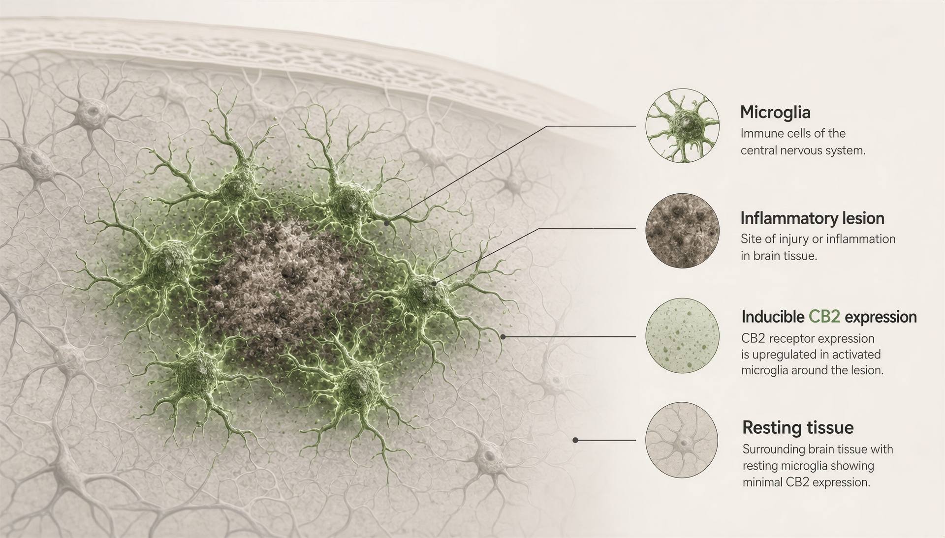
Microglia are the resident immune cells of the brain and spinal cord. In a healthy, unstimulated CNS, CB2 expression is generally low compared with immune organs like spleen. That low baseline is one reason older papers and textbooks often treated CB2 as effectively absent from the brain. But inflamed brain tissue is not a healthy resting baseline, and microglia are not passive bystanders. When activated by injury, infection, neurodegeneration, or chronic inflammatory signalling, microglia can upregulate CB2 quite markedly.
This finding has shown up across many disease models: multiple sclerosis, neuropathic pain, traumatic brain injury, Alzheimer’s disease, Parkinsonian models, and stroke, among others. The details differ, and not every reported increase is equally convincing. Still, the broad pattern has held up well enough that CB2 is now widely discussed as an inducible neuroimmune receptor. In these settings, CB2 is often detected in activated microglia clustered around lesions or areas of pathology rather than evenly distributed through normal brain parenchyma.
Why does that matter? Because neuroinflammation is not just “brain inflammation” in a vague sense. It alters synapses, neuronal survival, myelination, pain sensitivity, and disease progression. If CB2 expression rises with microglial activation, then cannabinoid signalling can affect CNS function without acting primarily through neuronal CB1. That helps explain why some cannabinoid effects in pain, neurodegeneration, and inflammatory models cannot be reduced to intoxication or classic psychoactivity.
The more controversial question is whether neurons themselves express meaningful amounts of CB2 in the CNS. Here the literature is mixed. Some studies have reported low-level CB2 mRNA or protein in subsets of neurons in the brainstem, hippocampus, cortex, or ventral tegmental area. Others have argued that many of those findings reflect antibody specificity problems, low-signal detection limits, species differences, or induction only under pathological conditions. Those are serious objections. CB2 research had a long period where weak tools produced overconfident localization claims.
A defensible position is this: constitutive neuronal CB2 expression in the healthy brain appears low and regionally restricted at most, not comparable to the dense and functionally dominant CB1 expression mapped across cortex, hippocampus, basal ganglia, and cerebellum. But low does not mean nonexistent, and inducible CNS expression under inflammatory or disease conditions is plausible and increasingly supported, especially in microglia and perhaps in selected neuronal populations depending on context.
That distinction matters for CBD discussions. CBD does not bind strongly to CB2 at physiologically typical concentrations, so claims that it works mainly by “activating CB2 in the brain” overstate the evidence. Still, any intervention that changes inflammatory signalling, endocannabinoid tone, adenosine signalling, TRP channel activity, or glial responses may intersect indirectly with CB2-linked pathways in neuroinflammatory states. The receptor is part of the network, not a one-step explanation.
Why “CB2 equals body” is too simple
The old CB1-brain/CB2-body split survives because it is memorable and partly true. CB1 is indeed the dominant cannabinoid receptor in the brain under baseline conditions, and CB2 is indeed much more prominent in immune cells and lymphoid tissue. As a first approximation, that is fine. As a biological model, it breaks down fast.
First, the brain is not immunologically separate from the rest of the body. Microglia are immune cells. Perivascular macrophages are immune cells. Infiltrating peripheral immune cells enter the CNS in disease. If CB2 tracks immune activation, then the brain can become a CB2-relevant organ whenever neuroinflammation is present. That is not a loophole. It is a central feature of the system.
Second, “body” is not one compartment. CB2 expression across peripheral tissues often reflects the density and state of resident or recruited immune cells rather than stable, high expression in every non-neural cell. Saying CB2 is “in the body” blurs the real pattern, which is enrichment in immune architecture and context-sensitive induction elsewhere.
Third, receptor distribution is dynamic. Expression changes with activation state, injury, cytokine milieu, developmental stage, and disease. A receptor map from healthy tissue can mislead if it is used to predict signalling during inflammation or degeneration. CB2 is one of the clearest examples of this principle in the ECS.
Fourth, pedagogical shortcuts distort drug claims. Once CB2 gets labeled the “body receptor,” it becomes easy to imply that compounds targeting it are nonpsychoactive, anti-inflammatory, and broadly therapeutic by default. The record does not support that kind of confidence. Receptor selectivity helps predict some effects, not all of them. Downstream biology still depends on timing, tissue, ligand bias, and pathology. The same lesson came from CB1 in reverse: pharmacologically manipulating the ECS can produce real clinical effects and real harm. Rimonabant, a CB1 inverse agonist, reduced weight in the RIO-Europe trial by 6.6 kg at one year versus 1.8 kg with placebo, then failed in practice because psychiatric adverse effects were serious enough to drive withdrawal. ECS signalling is powerful biology, not a wellness metaphor.
So the cleanest position is also the least catchy: CB2 is best understood as an immune-skewed, inflammation-responsive cannabinoid receptor with strong expression in B cells, NK cells, macrophages, spleen, tonsils, and related compartments, plus inducible relevance in the CNS, especially through microglia. That is more accurate than “CB2 equals body,” and accuracy matters here. Oversimplified receptor maps lead directly to oversimplified claims about what cannabinoids, including CBD, are likely to do.
The endogenous ligands: anandamide and 2-AG are not interchangeable
A lot of ECS explainers make a basic mistake: they treat anandamide and 2-arachidonoylglycerol, or 2-AG, as if they were two versions of the same internal cannabis-like signal. They are not. Both are endogenous lipids that can activate cannabinoid receptors, and both are produced on demand rather than stored in vesicles like classical neurotransmitters. But their chemistry, abundance, receptor efficacy, kinetics, and physiological jobs differ enough that collapsing them into one category hides how the system actually works.
That distinction matters for CBD discussions. If a compound shifts fatty acid amide hydrolase activity, alters anandamide tone, or changes TRPV1 signalling, that is not the same as changing 2-AG-mediated synaptic suppression. “Boosting endocannabinoids” sounds simple. It is not. The ECS is a lipid signalling network with division of labor, and AEA and 2-AG sit in different parts of that labor map.
Anandamide: synthesis, receptor activity, and naming
Anandamide was the first endocannabinoid identified. In 1992, William Devane, Lumír Hanuš, Raphael Mechoulam, and colleagues reported the isolation and characterization of arachidonoylethanolamide from porcine brain. They named it “anandamide” after ananda, the Sanskrit word for bliss, combined with the amide chemical suffix. The name helped it stick in public memory. The pharmacology is more complicated than the nickname suggests.
Chemically, anandamide is an N-acylethanolamine, often abbreviated AEA. It is generally formed from membrane phospholipid precursors, especially N-arachidonoyl phosphatidylethanolamine, through calcium-sensitive and enzyme-dependent pathways. The best-known route involves NAPE-PLD, N-acyl phosphatidylethanolamine phospholipase D, though that is not the only biosynthetic path. This already tells you something important: AEA is not a standing pool waiting in reserve. It is generated locally when cells need it.
Partial agonist A ligand that activates a receptor but does not produce the receptor's maximum possible response even when occupancy is high.
At cannabinoid receptors, AEA behaves mainly as a partial agonist, especially at CB1. That partial agonism separates it from 2-AG. AEA can activate CB1, but it does not usually drive the same maximal response that a full agonist can produce in the same system. Its effects depend heavily on receptor density, local synthesis, degradation rate, and what else is happening around the synapse. In tissues with dense CB1 expression, AEA can still have meaningful effects. Yet its signalling profile is often more selective and less quantitatively dominant than 2-AG.
AEA also refuses to stay inside the cannabinoid box. It interacts with targets outside CB1 and CB2, most notably TRPV1, the transient receptor potential vanilloid 1 channel that also responds to capsaicin. That matters because AEA can therefore influence pain signalling, inflammation, and sensory processing through routes that are not simply “cannabinoid receptor activation.” In some contexts, rising AEA can engage CB1-mediated inhibition; in others, TRPV1 activation may alter or even oppose expected cannabinoid-type effects. This is one reason simplistic language about “raising anandamide” often overstates therapeutic predictability.
Tissue levels of AEA are usually lower than those of 2-AG, especially in brain. It is present in nanomolar ranges where 2-AG often appears at much higher concentrations. Lower abundance does not mean unimportant. It means AEA likely functions more as a finely tuned signal than as the bulk workhorse ligand of fast retrograde cannabinoid transmission. Vincenzo Di Marzo and others have long emphasized that endocannabinoid signalling is context-dependent; AEA is one of the clearest examples of that principle.
Termination is also distinctive. AEA is primarily hydrolyzed by FAAH, fatty acid amide hydrolase, into arachidonic acid and ethanolamine. FAAH sits as a major checkpoint on AEA tone. If FAAH activity drops, AEA levels can rise. But even here, the biology resists simple summaries. FAAH inhibition does not just affect AEA; it can alter other fatty acid amides as well, which means the downstream physiology may reflect a broader lipid shift rather than a pure “anandamide increase.”
So AEA is not the endogenous equivalent of THC in any straightforward sense. It is shorter-lived, more locally constrained, only a partial agonist at CB1, and active at non-cannabinoid targets. That is a very different signalling style from a plant cannabinoid that enters the bloodstream, reaches multiple tissues, and persists far longer than the endogenous pulse it is often said to mimic.
2-AG: abundance, full agonism, and synaptic function
If AEA is the more famous endocannabinoid, 2-AG is often the more important one in day-to-day synaptic physiology. In 1995, groups led by Raphael Mechoulam and Tomoyuki Sugiura identified 2-arachidonoylglycerol as an endogenous ligand for cannabinoid receptors. That finding changed the picture of the ECS. It was no longer a system with one odd lipid messenger. It was a broader signalling architecture, and 2-AG turned out to be central to it.
2-AG is usually the quantitatively dominant endocannabinoid in the brain. Its tissue levels are commonly much higher than AEA levels, often by orders of magnitude depending on region and assay method. More than that, 2-AG acts as a full agonist at CB1 and CB2 in many experimental systems. That gives it a different functional profile from AEA. When 2-AG is synthesized at a synapse and reaches presynaptic CB1 receptors, it can strongly suppress neurotransmitter release.
This is where 2-AG becomes indispensable to understanding retrograde signalling. In many forms of short-term synaptic plasticity, postsynaptic depolarization or activation of certain Gq/11-coupled receptors raises intracellular calcium and triggers enzymatic production of 2-AG from diacylglycerol, mainly through diacylglycerol lipase alpha, DAGLα. The newly formed 2-AG then diffuses backward across the synaptic cleft and activates presynaptic CB1 receptors. The result is reduced release probability of GABA or glutamate.
Late-1990s and early-2000s electrophysiology work by researchers including Bradley Alger, Beat Lutz, Giovanni Marsicano, and Pablo Castillo helped define this process in functional terms. Depolarization-induced suppression of inhibition, DSI, and depolarization-induced suppression of excitation, DSE, are classic examples. In these cases, the postsynaptic neuron briefly tells the presynaptic neuron to quiet down. Endocannabinoids are the message. In many brain regions, 2-AG appears to be the dominant messenger carrying that message.
That role makes 2-AG less like a diffuse wellness molecule and more like a fast local regulator of circuit gain. It shapes how much inhibition or excitation gets through. It participates in stress responses, pain pathways, reward processing, learning, and memory. It can support both short-term and long-term plasticity depending on the circuit and timing. This is why “THC binds CB1” is such an incomplete explanation of cannabis action. The native ligand often controlling those CB1 receptors is 2-AG, released in tightly timed bursts at specific synapses, then shut down quickly.
Its degradation pathway reinforces that point. 2-AG is hydrolyzed primarily by monoacylglycerol lipase, MAGL. In a 2011 Nature Chemical Biology paper, Nomura and colleagues estimated that MAGL accounts for about 85% of 2-AG hydrolysis activity in mouse brain, with ABHD6 and ABHD12 contributing smaller fractions. That means 2-AG signalling is tightly regulated by a dedicated catabolic system. Turn MAGL down, and you do not just gently support ECS function; you can flood circuits with prolonged cannabinoid tone, alter eicosanoid metabolism, and potentially trigger receptor desensitization.
Compared with AEA, then, 2-AG is generally more abundant, often more efficacious at cannabinoid receptors, and more central to classic retrograde suppression of transmitter release. Calling both molecules “the body’s natural THC” is catchy but wrong. They operate on different scales and with different consequences.
Other endocannabinoid-related lipids and why they matter
Even the AEA-plus-2-AG story is incomplete. The ECS sits inside a larger lipid signalling environment that includes several endocannabinoid-related molecules. Some do not strongly activate CB1 or CB2 at all, yet they influence inflammation, feeding, pain, satiety, and receptor cross-talk. Ignoring them produces a cartoon version of the system.
Endocannabinoid-related lipids named in the article
- PEA
- Palmitoylethanolamide; discussed mainly for anti-inflammatory and analgesic effects, often through PPAR-α and mast-cell related pathways rather than strong direct CB1 agonism.
- OEA
- Oleoylethanolamide; linked more strongly to satiety, feeding regulation, and metabolic signalling, again with a major role for PPAR-α rather than direct cannabinoid receptor activation.
Two of the most important are palmitoylethanolamide, PEA, and oleoylethanolamide, OEA. Like AEA, they are N-acylethanolamines. They are produced from membrane lipid precursors and can be regulated by overlapping enzymatic machinery, including FAAH in some contexts. But they are not simply weaker copies of anandamide. Their pharmacology differs.
PEA has been studied mainly for anti-inflammatory and analgesic effects, often linked to PPAR-α signalling, mast-cell modulation, and indirect interactions with cannabinoid pathways rather than strong direct CB1 agonism. OEA is associated more strongly with satiety, feeding regulation, and metabolic signalling, again with a major role for PPAR-α rather than direct cannabinoid receptor activation. These compounds matter because manipulating FAAH or changing lipid precursor pools can shift several signalling molecules at once. A rise in AEA may come with altered PEA and OEA levels, and those changes can contribute to the observed biological effect.
This is one reason CBD pharmacology is still debated. CBD has low direct affinity for CB1 and CB2 at physiologically relevant concentrations, so claims that it “works by activating the ECS” are too neat. In some studies, CBD has been linked to altered anandamide signalling, possibly through FAAH-related mechanisms or transport-related effects, though the exact mechanism remains unsettled and may vary by model. If CBD changes fatty acid amide handling, the consequence may involve not just AEA but a family of related lipids. That is more plausible than the tidy idea that CBD simply raises one bliss molecule and restores balance.
Human evidence for broad entourage-effect claims is much thinner than common marketing language suggests. Limited evidence
The same caution applies to “entourage effect” rhetoric. Multi-compound interactions are pharmacologically plausible; that part is not controversial. What is controversial is how often those interactions have been demonstrated clearly in humans, at meaningful doses, with defined endpoints. The evidence is far thinner than marketing language has implied for years. Endocannabinoid-related lipids do interact. But plausible is not proven.
The broader lesson is straightforward: the endogenous cannabinoid system is not a two-key lock with THC as the spare key. It is a network of on-demand lipids, receptors, enzymes, and nearby signalling systems. AEA and 2-AG anchor that network, but they do not do the same job. AEA is lower-abundance, partial, and pharmacologically broader. 2-AG is the high-abundance full agonist that often carries fast retrograde synaptic signalling. Around them sits a wider family of bioactive lipids that can reshape the outcome. Any serious account of CBD, THC, or ECS-targeted therapy has to start there.
How endocannabinoid signals are made and shut down
The endocannabinoid system does not work like a warehouse with pre-packed messenger molecules waiting for release. It works more like a rapid-response lipid signalling network. That distinction matters. Classical neurotransmitters such as glutamate, GABA, dopamine, and serotonin are synthesized ahead of time, loaded into synaptic vesicles, and released in pulses when neurons fire. Endocannabinoids are different. Anandamide (AEA) and 2-arachidonoylglycerol (2-AG) are usually made on demand from membrane lipids, act over very short distances, and are then dismantled quickly. Their short life is part of their job.
This is one reason popular shorthand like “CBD boosts the ECS” or “THC activates the body’s natural cannabis system” is misleading. The endogenous system is tightly timed, highly local, and enzymatically shut off within moments. Phytocannabinoids enter that network from the outside and often behave very differently in duration, spread, and receptor occupancy.
On-demand synthesis from membrane lipids
Endocannabinoids are not stored in synaptic vesicles. Neurons and other cells synthesize them when needed from phospholipid precursors embedded in cell membranes. That on-demand feature was one of the major conceptual shifts that followed the discovery of CB1 by Matsuda et al. in 1990, anandamide by Devane et al. in 1992, CB2 by Munro et al. in 1993, and 2-AG as an endocannabinoid in 1995 by Mechoulam and colleagues and independently by Sugiura’s group.
In the brain, the best-characterized trigger is a rise in postsynaptic intracellular calcium, often combined with activation of Gq/11-coupled receptors. When the postsynaptic neuron is strongly depolarized, or when certain metabotropic receptors are activated, enzymes in the membrane start cutting endocannabinoid precursors into active signalling lipids. The result is a messenger that can move backward across the synapse and tell the presynaptic terminal to release less neurotransmitter. That is retrograde signalling.
For anandamide, the biochemistry is more complicated than many diagrams suggest, but the non-specialist version is manageable. AEA is generated from membrane phospholipids that have first been converted into N-acyl phosphatidylethanolamines, often shortened to NAPEs. One major route then uses the enzyme NAPE-PLD, or N-acyl phosphatidylethanolamine-selective phospholipase D, to produce anandamide from those NAPE precursors. NAPE-PLD is not the whole story. Alternative pathways exist, and different tissues may lean on different enzymatic routes. That complexity is one reason AEA biology can look inconsistent across experiments.
2-AG follows a somewhat clearer path. Its immediate precursor is diacylglycerol, or DAG, a lipid intermediate generated in membranes after phospholipase C cuts phosphoinositides. DAG is then converted to 2-AG by diacylglycerol lipase, usually DAGL-alpha in neurons and DAGL-beta in some other cell types. If you want the simple picture, it is this: neuronal activity changes membrane lipid chemistry, and that chemistry is rapidly transformed into an endocannabinoid pulse.
The location of the synthetic machinery helps explain the direction of signalling. In many central synapses, DAGL-alpha is enriched postsynaptically, while CB1 receptors are concentrated presynaptically. That anatomical arrangement supports the classic scheme worked out in late-1990s and early-2000s electrophysiology by researchers including Bradley Alger, Beat Lutz, Giovanni Marsicano, Daniele Piomelli, Stella, and Castillo: a postsynaptic neuron becomes active, makes endocannabinoids on demand, sends them backward across the synaptic cleft, and suppresses transmitter release from the presynaptic terminal.
This can happen over seconds, as in depolarization-induced suppression of inhibition (DSI) or depolarization-induced suppression of excitation (DSE), where endocannabinoids transiently reduce release of GABA or glutamate. It can also contribute to longer-lasting forms of synaptic plasticity. The point is not just that endocannabinoids exist. It is that their synthesis is yoked to local activity. They are event-driven signals.
AEA and 2-AG are not interchangeable here. AEA is usually present at lower concentrations and often behaves as a partial agonist at CB1. 2-AG is generally the quantitatively dominant endocannabinoid in the brain and often acts as a full agonist at CB1 and CB2 in many assay systems. In practical terms, 2-AG is often the workhorse of fast synaptic retrograde signalling, while AEA may have more selective or context-dependent roles. There is overlap, but flattening them into “the body’s natural cannabinoids” misses real functional differences.
FAAH and anandamide degradation
Once anandamide has done its job, the signal has to stop. That stop signal is not an afterthought. It is part of the design.
| Enzyme | Main substrate or role | Article note |
|---|---|---|
| FAAH | Anandamide (AEA) hydrolysis | Major checkpoint on AEA tone |
| MAGL | 2-AG hydrolysis | About 85% of 2-AG hydrolysis activity in mouse brain |
| ABHD6 | Local 2-AG regulation | Smaller contributor, often near postsynaptic membranes |
| ABHD12 | 2-AG-related lipid turnover | Smaller contributor with neuroimmune relevance |
The main enzyme responsible for anandamide degradation is FAAH, fatty acid amide hydrolase. FAAH sits largely on intracellular membranes, especially the endoplasmic reticulum, and hydrolyzes AEA into arachidonic acid and ethanolamine. Because AEA is lipophilic, it diffuses through membranes rather than behaving like a water-soluble neurotransmitter in an open extracellular pool. After uptake or membrane partitioning, FAAH clears it quickly.
That rapid hydrolysis keeps anandamide signalling brief and spatially restricted. Without fast breakdown, AEA would spread farther, last longer, and blur the distinction between active and inactive synapses. In that sense, FAAH is not just housekeeping. It shapes the message itself by controlling how big the signal becomes and how long it can influence nearby receptors.
This is one place where external cannabinoids diverge sharply from endogenous ones. THC is not rapidly cleared by FAAH. It can occupy CB1 receptors for much longer and in many more brain regions at once than a naturally generated AEA pulse would. So even when THC and AEA hit the same receptor, they do not create the same physiological event. Timing matters. Locality matters. Enzymatic shutdown matters.
FAAH became an obvious drug target for that reason. In theory, inhibiting FAAH should raise anandamide only where and when it is being produced, offering a subtler way to amplify endocannabinoid tone than directly stimulating CB1 receptors. That idea was attractive, especially after the psychiatric problems caused by rimonabant, the CB1 inverse agonist marketed for obesity before being withdrawn. But the story is a warning against simplistic “boost the ECS” thinking. The 2016 BIA 10-2474 phase 1 trial in France, involving a FAAH inhibitor, caused severe neurotoxicity and one death. The exact mechanism remains debated and likely involved off-target effects rather than FAAH inhibition alone, but the broader lesson stands: manipulating endocannabinoid shutdown is pharmacology with real risk, not gentle system support.
CBD is sometimes described as a FAAH inhibitor. That claim needs restraint. In some preclinical or in vitro contexts, CBD can affect FAAH-related pathways or anandamide levels, but it is not accurate to present its human effects as a straightforward FAAH-blocking mechanism. Its pharmacology is broader and messier than that.
MAGL, ABHD6, and ABHD12 in 2-AG clearance
If FAAH is the main off-switch for anandamide, monoacylglycerol lipase, or MAGL, is the dominant off-switch for 2-AG. This is one of the clearest quantitative findings in ECS biochemistry. Nomura et al., writing in Nature Chemical Biology in 2011, estimated that MAGL accounts for about 85% of 2-AG hydrolysis activity in mouse brain. The rest is handled largely by two serine hydrolases: ABHD6 and ABHD12.
That division of labor matters because 2-AG is usually the major endocannabinoid signal in the central nervous system. If you want to understand how cannabinoid signalling is terminated in the brain, you have to understand MAGL first.
MAGL is found primarily in presynaptic compartments in many neuronal circuits, a fitting location given that 2-AG often acts on presynaptic CB1 receptors after being synthesized postsynaptically. A common sequence is: postsynaptic activity drives 2-AG production via DAGL, 2-AG diffuses retrogradely to the presynaptic terminal, CB1 activation suppresses neurotransmitter release, and MAGL then hydrolyzes 2-AG to end the signal. The signal is therefore built around both directionality and timed destruction.
ABHD6 and ABHD12 are smaller contributors in bulk hydrolysis terms, but “smaller” does not mean trivial. ABHD6 is often associated with postsynaptic membranes and may regulate local 2-AG availability close to its site of synthesis, effectively shaping the signal before it fully develops. ABHD12 appears to contribute more in microglia and other cell types, with broader implications for neuroimmune signalling. Mutations in ABHD12 cause the rare neurodegenerative disorder PHARC, which includes polyneuropathy, hearing loss, ataxia, retinitis pigmentosa, and cataract, a reminder that lipid hydrolases in this pathway are not minor accessories.
Fast 2-AG breakdown also has another consequence: it links cannabinoid signalling to arachidonic acid metabolism. Because MAGL hydrolysis yields arachidonic acid and glycerol, MAGL sits at an interface between endocannabinoid signalling and eicosanoid biology. Inflammatory consequences can follow. Block MAGL hard enough, and you are not only changing CB1 and CB2 signalling. You may also be reshaping downstream lipid mediator pools.
So shutdown is not cleanup after the interesting part. Shutdown is the interesting part. It determines whether an endocannabinoid signal remains synapse-specific or becomes diffuse, whether it lasts milliseconds or minutes, and whether pharmacological intervention produces subtle modulation or receptor overdrive. That is the frame to keep in mind when comparing endogenous cannabinoids with plant cannabinoids. The body’s own signals are made late, nearby, and briefly. THC, CBD, and other phytocannabinoids arrive early, spread widely, and ignore much of the built-in timing logic.
Retrograde synaptic signalling: the mechanism that made the ECS famous
The endocannabinoid system became a serious neuroscience story when researchers showed that its main synaptic trick runs backward. In the standard textbook direction, presynaptic terminals release neurotransmitter and postsynaptic cells respond. Endocannabinoids often reverse that flow of information. A postsynaptic neuron that has just been strongly activated can synthesize its own lipid messengers on demand, release them into the synaptic space, and tell the presynaptic terminal to quiet down. That is retrograde signalling.
This is the point many cannabis explainers miss. The ECS is not just “THC binds CB1.” It is a timing-sensitive feedback network built from membrane lipids, calcium signals, G-protein-coupled receptors, and fast enzymatic shutoff. THC can plug into that machinery, but it does not reproduce its normal rhythm very well.
Late-1990s and early-2000s electrophysiology made this mechanism hard to ignore. Work from Bradley Alger, Beat Lutz, Giovanni Marsicano, Vincenzo Di Marzo, Ken Mackie, George Kunos, and others showed that endocannabinoids could suppress transmitter release across many brain regions. Daniel Castillo and colleagues then helped establish how this system contributes not only to brief synaptic silencing, but to durable forms of plasticity. The result was a major shift in how synapses were understood: postsynaptic cells are not passive receivers. They vote back.

From postsynaptic calcium rise to presynaptic CB1 activation
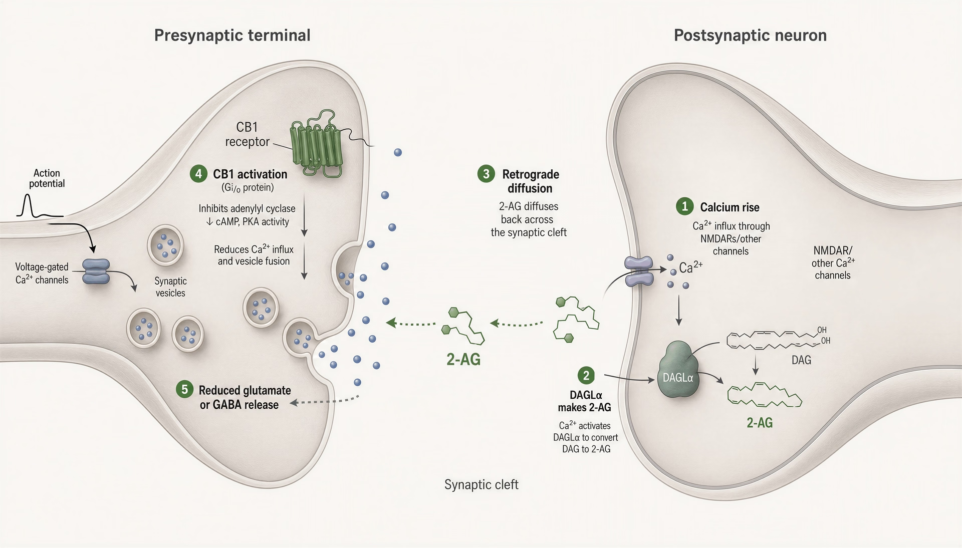
The sequence begins in the postsynaptic neuron. Strong depolarization, intense synaptic input, or activation of certain Gq/11-coupled receptors raises intracellular calcium. That calcium rise is the trigger. It activates enzymatic pathways that build endocannabinoids from membrane phospholipid precursors rather than releasing them from pre-stored vesicles.
Two endocannabinoids matter most here: anandamide (AEA) and 2-arachidonoylglycerol (2-AG). They are not interchangeable. In most fast retrograde synaptic signalling in the brain, 2-AG appears to do most of the work. It is usually present in much higher amounts than anandamide and acts as a full agonist at CB1 in many systems. Anandamide is often lower in concentration, shorter-lived in some contexts, and behaves as a partial agonist at CB1. That difference matters because synaptic suppression depends on amplitude, timing, and receptor occupancy, not on a vague idea of “more ECS tone.”
For 2-AG, the usual pathway runs through phospholipase C and diacylglycerol lipase, especially DAGL-alpha in many excitatory synapses. Membrane lipids are converted into diacylglycerol, then into 2-AG. Anandamide is produced through different routes, often involving NAPE-derived intermediates. The key principle is on-demand synthesis. Endocannabinoids are made when needed, near the synapse that needs them.
Once produced, these lipids diffuse out of the postsynaptic membrane and move across the synaptic cleft. No vesicle fusion is required. They then bind CB1 receptors on the presynaptic terminal. CB1, cloned by Matsuda et al. in Nature in 1990, is one of the most abundant GPCRs in the brain, especially in cortex, hippocampus, basal ganglia, cerebellum, and several limbic areas. It is positioned perfectly for this job.
CB1 is Gi/o-coupled. When activated, it suppresses adenylyl cyclase, reduces calcium influx through voltage-gated calcium channels, and can increase potassium conductance through inwardly rectifying channels. The practical effect at the terminal is simple: vesicle release becomes less likely. That means less glutamate if the presynaptic neuron is excitatory, or less GABA if it is inhibitory.
Then the signal is shut down. Fast. Anandamide is primarily degraded by FAAH. Brain 2-AG is mostly hydrolyzed by MAGL; Nomura et al. reported in 2011 that MAGL accounts for about 85% of 2-AG hydrolysis activity in mouse brain, with ABHD6 and ABHD12 handling smaller fractions. This rapid termination is part of the design. Endocannabinoids are local feedback signals, not meant to soak the entire brain for hours.
Depolarization-induced suppression of inhibition and excitation
The two classic demonstrations of retrograde cannabinoid signalling are DSI and DSE: depolarization-induced suppression of inhibition and depolarization-induced suppression of excitation.
In DSI, a postsynaptic neuron depolarizes, intracellular calcium rises, and endocannabinoids are released backward onto CB1-expressing GABAergic terminals. GABA release falls for a short period, often seconds to tens of seconds depending on the preparation. The postsynaptic cell is temporarily less inhibited. It has, in effect, loosened the brake.
In DSE, the same basic logic applies, but now the target is an excitatory glutamatergic terminal. Endocannabinoid release suppresses glutamate release. The accelerator is eased off.
These phenomena were first characterized in brain-slice electrophysiology in regions such as the hippocampus and cerebellum, then extended to many other circuits. They mattered because they showed that endocannabinoid signalling was not exotic or rare. It was woven into ordinary synaptic control.
The exact pattern depends on where CB1 is expressed. In some circuits, CB1 is especially dense on particular classes of inhibitory interneurons, making DSI prominent. In others, glutamatergic terminals also show cannabinoid sensitivity, supporting DSE. Receptor distribution is not uniform, and that unevenness is one reason global cannabinoid exposure produces mixed effects. A single drug can suppress inhibition in one microcircuit and suppress excitation in another.
That is why the phrase “cannabis calms the nervous system” is too sloppy to be useful. Sometimes CB1 activation reduces excitatory drive and dampens network activity. Sometimes it suppresses inhibition and disinhibits neurons. Sometimes both happen in parallel across different cell types. The net result depends on region, cell identity, firing state, receptor density, and dose.
These local feedback loops help explain several behavioral effects that otherwise look unrelated. In pain pathways, endocannabinoid-mediated suppression can reduce nociceptive transmission and shape descending pain control. In amygdala-prefrontal circuits, it can support fear extinction by allowing outdated threat associations to weaken under the right conditions; Giovanni Marsicano and colleagues provided influential evidence for this in Nature in 2002, showing that CB1 signalling was required for extinction of aversive memories in mice. In reward circuits, cannabinoid modulation changes inhibitory and excitatory balance in the ventral tegmental area and nucleus accumbens, altering dopamine-related signaling. In hippocampal networks, it affects oscillations, information flow, and memory encoding.
Short-term versus long-term synaptic plasticity
DSI and DSE are short-term plasticity. They last seconds to minutes. They act like a rapid feedback brake, allowing an active postsynaptic neuron to tune incoming drive in real time. That alone would make the ECS important. But the system also participates in longer-lasting synaptic change.
Endocannabinoid-dependent long-term depression, usually called eCB-LTD, has been described in the striatum, cortex, hippocampus, nucleus accumbens, amygdala, and cerebellum. Here the same basic ingredients recur: postsynaptic activity, endocannabinoid synthesis, retrograde activation of presynaptic CB1, and a sustained reduction in release probability. The difference is persistence. Instead of transient suppression, repeated or patterned activity can push the synapse into a lower-output state that lasts much longer.
That matters for learning. In corticostriatal pathways, eCB-LTD is tied to habit formation, action selection, and motor learning. In the amygdala and medial prefrontal cortex, it influences emotional learning and extinction. In the hippocampus, it can shape the filtering of information and the threshold for memory formation. In addiction-related circuitry, repeated drug exposure can alter endocannabinoid plasticity itself, changing how reward and cue learning are regulated.
This is also where THC begins to look less like a clean substitute and more like a system-wide perturbation. Endogenous signalling is generated on demand, confined to active synapses, and terminated quickly by FAAH and MAGL. THC arrives from outside, reaches many CB1-rich regions at once, and lingers far longer than a normal retrograde burst. It does not wait for a specific postsynaptic calcium event. It does not respect synapse-level boundaries. So while THC can mimic part of the retrograde signal by activating CB1, it can also override the logic of the circuit.
That distinction helps explain both therapeutic promise and side effects. A well-timed endogenous endocannabinoid signal may sharpen circuit control. Broad CB1 activation by THC can instead impair working memory, disrupt temporal coding, alter cerebellar processing, and distort reward learning. The same receptor is involved. The pattern of activation is not.
CBD is a different case. It has low direct affinity for CB1 and CB2 at typical physiological concentrations and does not simply reproduce retrograde cannabinoid transmission. Claims that CBD “supports the ECS” are usually too vague to mean much. Its actions appear to involve a mixed pharmacology that may include TRPV1, 5-HT1A, adenosine-related signaling, ion channels, and possibly context-dependent effects on endocannabinoid tone. That is pharmacologically interesting, but it is not the same as saying CBD neatly boosts the brain’s native retrograde feedback system.
So the famous mechanism is not sedation. It is precision control. Endocannabinoid retrograde signalling lets active neurons regulate the inputs they receive, moment by moment and synapse by synapse. That is a much more exact account of how the ECS shapes neural function than the popular claim that cannabinoids simply “relax” the brain.
The ECS across major body systems
The endocannabinoid system is often described as a “balance” network, but that shorthand can mislead. The ECS does not patrol the body looking for anything out of range and then neatly restore health. It adjusts signalling thresholds, often in short-lived, local, context-dependent ways. In one tissue that may dampen neurotransmitter release; in another it may restrain cytokine production; in a third it may alter gut motility or hypothalamic feeding signals. Sometimes those adjustments are adaptive. Sometimes they are not. And when phytocannabinoids such as THC enter the picture, the pattern no longer resembles the tight timing of endogenous ligands made on demand and broken down quickly by FAAH or MAGL.
Evidence strength also varies sharply by system. The strongest case for ECS importance is in nervous system signalling and in immune and inflammatory regulation. Appetite and emesis control are also well supported. Claims about endocrine disease, metabolism, fertility, or “supporting homeostasis” are much softer and often oversold.
Nervous system: pain, stress, memory, appetite, reward, sleep
The nervous system is where ECS biology is most established. That starts with receptor distribution. CB1, cloned by Matsuda et al. in Nature in 1990, is one of the most abundant G protein-coupled receptors in the brain, with high expression in cortex, hippocampus, basal ganglia, cerebellum, and limbic circuitry. That map matters. It predicts the real-world effects of THC rather well: altered memory, attention, motor coordination, reward processing, appetite, and time perception. It also helps explain what cannabis usually does not do. CB1 expression is sparse in the medullary cardiorespiratory centers that drive breathing, which is one reason cannabinoid overdose does not produce the same fatal respiratory depression pattern seen with opioids.
Mechanistically, the ECS is built for synaptic fine-tuning. Late-1990s and early-2000s electrophysiology work by Bradley Alger, Patrice Stella, Pablo Castillo, and others clarified the basic motif: postsynaptic activity raises intracellular calcium or activates certain GPCR pathways, which triggers synthesis of endocannabinoids from membrane lipid precursors. Those endocannabinoids then travel backward across the synapse and activate presynaptic CB1 receptors. The result is reduced probability of neurotransmitter release, whether inhibitory GABA or excitatory glutamate. This is the basis of depolarization-induced suppression of inhibition and excitation, and of several forms of short- and long-term synaptic plasticity.
Pain regulation is one of the clearest system-level consequences. CB1 receptors are present along nociceptive pathways in peripheral terminals, spinal cord dorsal horn, and supraspinal pain circuits. CB2 has a larger role in inflammatory and immune-linked pain states. Endocannabinoids can reduce transmitter release in pain pathways and alter descending pain control. Human therapeutic evidence is imperfect but meaningful. The 2017 National Academies report judged there to be substantial evidence that cannabis or cannabinoids are effective for chronic pain in adults. That does not mean the ECS is a universal analgesic switch, and it does not settle which products, doses, ratios, or routes are optimal. Still, pain is one of the areas where ECS modulation has moved beyond speculation.
Stress signalling is also deeply entangled with endocannabinoids. AEA and 2-AG participate in feedback control within the amygdala, prefrontal cortex, hippocampus, and hypothalamus. Acute stress can lower AEA tone in some circuits and shift 2-AG signalling later in the response, with consequences for anxiety, arousal, and recovery. Giovanni Marsicano and colleagues showed in the early 2000s that CB1 signalling is involved in extinction of aversive memories in animal models, a finding that helped drive interest in cannabinoids for trauma-related symptoms. But translation has been uneven. There is no clean, universal rule that “more cannabinoid signalling means less anxiety.” Low doses of THC may reduce anxiety in some people and settings; higher doses often do the opposite. CBD’s anxiolytic literature is intriguing but mechanistically messy and not reducible to direct CB1 or CB2 activation.
Memory effects are a place where public summaries often get the direction right but the biology wrong. THC disrupts short-term memory largely because CB1 is dense in the hippocampus and cortical networks involved in encoding and retrieval. Endogenous signalling in those circuits is normally brief and spatially restricted. THC is neither. It lingers, reaches multiple regions at once, and can override endogenous timing. That distinction matters. The ECS supports synaptic plasticity; exogenous cannabinoids can distort it. Chronic heavy exposure, especially during adolescence, appears more likely to impair learning and memory than to “normalize” them.
Appetite regulation is one of the oldest and strongest physiological observations linked to the ECS. CB1 signalling in hypothalamic and mesolimbic pathways increases feeding drive and the salience of palatable food. Endocannabinoids rise during fasting in ways that fit this role. THC can mimic that effect. The anti-obesity drug rimonabant, a CB1 inverse agonist, offered proof by reversal: block CB1 and weight falls. In the 2005 RIO-Europe trial published in Lancet, Van Gaal et al. reported one-year weight loss of 6.6 kg with rimonabant 20 mg versus 1.8 kg with placebo. But the drug was withdrawn because psychiatric adverse effects, including depression and anxiety, were too serious. That episode is one of the clearest warnings in ECS pharmacology. Manipulating this system can produce real therapeutic effects. It can also produce real harm.
Reward and reinforcement sit in the same category. CB1 modulates dopamine-linked circuitry in the ventral tegmental area and nucleus accumbens, though often indirectly through effects on GABAergic and glutamatergic inputs rather than by simply “flooding the brain with dopamine.” THC can increase salience and reinforcement, which is part of why repeated use becomes compulsive in a subset of users. NIDA notes that about 3 in 10 cannabis users may develop cannabis use disorder. That statistic should not be inflated into a claim that all cannabinoid signalling is addictive, but it does contradict the lazy view that ECS-active compounds are inherently self-limiting or gentle.
Sleep is another mixed but legitimate ECS domain. Endocannabinoids fluctuate with circadian and vigilance states, and CB1 signalling influences sleep onset, architecture, and arousal systems. THC often shortens sleep latency in the short term, while chronic use and withdrawal can disrupt sleep continuity and dreaming. CBD is even less straightforward: some studies suggest alerting effects at certain doses and sedating effects at others. The main point is that the ECS shapes sleep regulation, but “better sleep” is not an automatic outcome of cannabinoid exposure.
Immune and inflammatory signalling
If CB1 is the dominant neural receptor, CB2 is the major immune-facing receptor, though the simplistic “CB1 equals brain, CB2 equals body” formula is not good enough. Munro et al. cloned CB2 in 1993, and subsequent work showed strong expression across immune cells and tissues, including B cells, macrophages, monocytes, and related lineages. Under inflammatory or pathological conditions, low-level CB2 expression in parts of the nervous system may increase, but claims about broad constitutive neuronal CB2 expression remain contested and highly context-sensitive.
Functionally, ECS signalling in immunity tends to regulate tone rather than act as an on/off switch. CB2 activation often reduces pro-inflammatory cytokine release, immune-cell migration, antigen presentation, or other features of activated immune responses. CB1 can also influence neuroinflammation and peripheral inflammatory signalling, though its role is more complicated because it intersects with neural regulation of inflammation as well as direct receptor actions in some tissues.
This is one of the areas where “homeostasis” language is most tempting and most prone to abuse. Inflammation is not inherently bad. It is a defensive program. The ECS can restrain excessive inflammatory signalling, which may protect tissue, but excessive suppression can also be maladaptive depending on the infection, injury, or disease state. Context decides.
The strongest evidence here is preclinical. In animal models, raising endocannabinoid tone or activating CB2 can reduce inflammatory markers in arthritis, colitis, neuropathic pain, and neuroinflammation paradigms. Human evidence exists, but it is much patchier than marketing copy suggests. For multiple sclerosis, the best-supported cannabinoid effect is not broad immune correction but symptom relief, especially spasticity and pain. Nabiximols has evidence in that setting in some jurisdictions. In inflammatory bowel disease, mechanistic plausibility is high because the gut is rich in ECS signalling elements, yet controlled clinical data remain mixed and do not justify sweeping claims of disease modification.
CBD is frequently placed in this immune section as though its role were settled. It is not. CBD has low direct affinity for CB1 and CB2 at physiologically relevant concentrations and likely acts through a spread of targets that may include TRPV1, 5-HT1A, PPAR-gamma, adenosine-related signalling, and ion channels, with FAAH-related effects in some contexts. Anti-inflammatory effects are plausible and often seen in vitro or in animal work, but direct clinical translation varies by condition. The same goes for CBG and lesser-studied cannabinoids: pharmacological plausibility is not the same thing as demonstrated patient benefit.
Endocrine, digestive, metabolic, and reproductive functions
This is where careful wording matters most. The ECS clearly participates in hypothalamic regulation, gut function, energy balance, and reproductive physiology. What is much less clear is how often targeting it improves disease outcomes without unacceptable tradeoffs.
In the endocrine sphere, the hypothalamus is the key hub. CB1 signalling intersects with circuits controlling appetite, stress hormone release, and neuroendocrine output. Endocannabinoids modulate hypothalamic-pituitary-adrenal axis responsivity, and interactions with glucocorticoid feedback are well described. But the literature is not consistent enough to support broad claims that cannabinoids “balance hormones.” They alter endocrine signalling. That is not the same thing.
Metabolic research is similarly mixed. CB1 signalling promotes feeding and lipogenesis in several contexts; peripheral CB1 activity has been linked to adiposity, insulin resistance, and dyslipidemia in animal and human studies. The rimonabant story showed that blocking CB1 can improve weight and some cardiometabolic markers, but it also showed why a mechanistically correct idea can fail clinically. There is ongoing interest in separating central from peripheral CB1 effects to avoid psychiatric toxicity, yet no simple therapeutic template has emerged. Claims that CBD improves insulin sensitivity or “resets metabolism” remain ahead of decisive human evidence.
The digestive tract has a better-defined ECS role. CB1 and CB2, along with endocannabinoid-metabolizing enzymes, are present in enteric neurons, epithelial cells, and immune components of the gut. Endocannabinoid signalling can slow gastrointestinal motility, affect secretion, and alter visceral sensation. Antiemetic effects are among the strongest clinically supported cannabinoid actions. The 2017 National Academies report found substantial evidence for cannabis or cannabinoids in chemotherapy-induced nausea and vomiting, and that fits older pharmacology involving brainstem and vagal emesis circuits. Here again, though, the system is not uniformly protective. Chronic heavy THC exposure can contribute to cannabinoid hyperemesis syndrome, a paradox that should end any simplistic claim that cannabinoids always normalize nausea pathways.
Interest in intestinal permeability and gut barrier function is real, especially because inflammatory states can alter ECS components in the intestine. Some preclinical models suggest barrier-protective effects under certain conditions. Human evidence is still preliminary and heterogeneous. It is fair to say the ECS is involved in gut barrier regulation. It is not fair to say cannabinoids reliably repair “leaky gut.”
Reproductive effects are even more reason for restraint. The ECS is active in gonads, implantation biology, placental function, and sperm physiology. AEA signalling appears to matter in implantation timing and embryo transport, while altered ECS tone has been linked to disrupted fertility in animal models and some human observational work. In males, heavy cannabis exposure has been associated in some studies with changes in sperm parameters and reproductive hormones, though findings are not perfectly consistent and confounding is common. The broad takeaway is modest but important: the ECS is part of reproductive physiology, and sustained external cannabinoid exposure can interfere with it. That is a stronger statement than many casual summaries make, and it is better supported than claims of reproductive benefit.
Across all of these systems, the central lesson is the same. The ECS is a real signalling network distributed through the brain, immune organs, gut, endocrine axes, and reproductive tissues. It helps organisms adjust to internal and external demands. But adjustment is not rescue, and perturbation is not therapy by default. Cannabis-derived compounds can tap into this network, sometimes usefully, sometimes bluntly, and sometimes in ways that expose how tightly tuned endogenous cannabinoid signalling normally is.
How phytocannabinoids interact with the ECS
The endocannabinoid system did not evolve so humans could respond to cannabis. It is an endogenous lipid signalling network that was discovered partly because THC pointed researchers toward it. That historical accident still distorts public explanations. Many articles imply that phytocannabinoids simply “fit” the ECS the way a key fits a lock. That is too neat. Endocannabinoids such as anandamide (AEA) and 2-arachidonoylglycerol (2-AG) are made on demand from membrane lipids, released locally, and then shut down quickly by enzymes such as FAAH and MAGL. Plant cannabinoids arrive from outside, often in much larger doses, by inhalation or ingestion, with very different pharmacokinetics. They do not just join the system. They perturb it.
That distinction matters. Endogenous ligands and phytocannabinoids differ in receptor efficacy, tissue exposure, timing, metabolism, and persistence. AEA and 2-AG are usually brief, spatially constrained signals. THC can bathe broad receptor populations at once and keep them occupied far longer than a physiological retrograde signal would. CBD is different again: weak direct binding at CB1 and CB2, but a broad and still partly unresolved pharmacology across non-cannabinoid targets. Minor cannabinoids complicate the picture further, though receptor charts often make them look more settled than they are.
THC as a partial agonist that can override endogenous timing
THC is often described as “the compound that binds CB1.” True, but incomplete. Mechanistically, delta-9-tetrahydrocannabinol is a partial agonist at both CB1 and CB2 receptors. “Partial” means it does not produce the maximal receptor response that a full agonist can produce, even when it occupies the receptor. That already separates it from the simplistic idea that THC just turns the ECS on. In many systems, 2-AG behaves as a higher-efficacy endogenous signal at CB1 and CB2 than THC does, and AEA has its own profile as a partial agonist. THC is therefore not a perfect stand-in for either endogenous ligand. It is a partial mimic.
The bigger issue is timing. Endocannabinoids are usually synthesized on demand in response to local activity. In the classic retrograde model worked out across late-1990s and early-2000s electrophysiology, postsynaptic activity raises calcium or triggers GPCR signalling, leading to production of AEA or 2-AG. These lipids travel backward across the synapse and activate presynaptic CB1 receptors, reducing release probability for glutamate or GABA. Then they are degraded. The signal is brief and targeted.
THC does not respect that timing logic. After inhalation, it reaches the brain quickly and distributes across CB1-rich regions including cortex, hippocampus, basal ganglia, cerebellum, and limbic circuits. After oral use, onset is slower and effects are often more prolonged, in part because of first-pass metabolism and formation of active 11-hydroxy-THC. Either way, the signal is exogenous, diffuse, and untethered from the local demand signals that normally generate endocannabinoids. THC can activate receptors on many terminals at once regardless of whether a given synapse “asked” for endocannabinoid feedback.
That is why calling THC a “natural fit” misses the point. It can override endogenous signalling rhythms. It may suppress neurotransmitter release where short-lived retrograde control would normally be absent, extend signalling where endogenous cannabinoids would already have been cleared, and alter network oscillations in circuits involved in memory, salience, motor control, and reward. The psychoactive effects of THC are not evidence that it neatly restores homeostasis. They are evidence that broad CB1 engagement changes information processing across distributed neural systems.
Persistence matters too. Endocannabinoids are terminated rapidly. AEA is mainly degraded by FAAH. About 85% of brain 2-AG hydrolysis is attributed to MAGL, with smaller contributions from ABHD6 and ABHD12, as shown by Nomura and colleagues in 2011. THC is not terminated by those same local shutdown mechanisms in the same way or on the same timescale. It is metabolized mainly in the liver, and its tissue distribution is shaped by lipophilicity, route of administration, repeated exposure, and accumulation in fat. That is a very different kinetic regime from an on-demand synaptic lipid messenger.
Repeated THC exposure adds another layer: receptor adaptation. CB1 receptors can desensitize and internalize after sustained agonist exposure, with region-specific differences. That helps explain tolerance and some withdrawal phenomena. If roughly 3 in 10 people who use cannabis develop cannabis use disorder, as NIDA has estimated, the answer cannot be “because the ECS likes cannabinoids.” The better explanation is that repeated exogenous perturbation of reward, stress, and habit circuitry can produce maladaptive neuroadaptation.
CBD: low direct receptor affinity, broad indirect pharmacology
CBD is often presented as the tidy opposite of THC: non-intoxicating, ECS-supporting, and straightforwardly therapeutic. The evidence does not support that simple story. Cannabidiol has low direct affinity for CB1 and CB2 at physiologically relevant concentrations compared with THC and many synthetic ligands. It is not best understood as a classic agonist at either receptor. If CBD has ECS effects, many appear to be indirect, context-dependent, and spread across several molecular systems.
One proposed mechanism is negative allosteric modulation at CB1. In plain terms, CBD may alter the receptor’s shape in a way that changes how other ligands signal through it. This has been reported in experimental systems and is pharmacologically plausible, but the size and in vivo relevance of that effect across doses and tissues are still debated. It is safer to say that CBD may modulate CB1 signalling rather than directly drive it.
FAAH is another common talking point. Some popular explainers claim CBD “raises anandamide” by blocking FAAH. That may happen in some contexts, but the picture is mixed. In vitro findings vary by system, concentration, and assay conditions, and CBD does not behave like a clean, potent FAAH inhibitor in the way dedicated experimental compounds do. Human evidence is suggestive in some settings but not decisive enough to reduce CBD to “a FAAH blocker.” Claims that CBD straightforwardly boosts the body’s own cannabinoids go beyond the evidence.
What is clearer is that CBD has broad polypharmacology. Targets proposed in preclinical and translational work include TRPV1 channels, 5-HT1A signalling, adenosine-related pathways, PPAR-gamma, GPR55, and several ion channels. Some of these interactions may help explain why CBD does not map neatly onto THC. Its anticonvulsant effects, for example, are unlikely to be explained by simple CB1 or CB2 agonism. That point is backed by clinical data. In Dravet syndrome, Devinsky et al. reported in the New England Journal of Medicine in 2017 that cannabidiol reduced convulsive seizure frequency by 43.9% versus 21.8% with placebo. In Lennox-Gastaut syndrome, Thiele et al. reported in 2018 that drop-seizure frequency fell 41.9% in the 20 mg/kg/day CBD group versus 17.2% with placebo. Those are real effects, but they come from a purified pharmaceutical product, Epidiolex, under controlled dosing and monitoring. They do not validate every broad claim attached to retail CBD.[8] Cannabidiol (CBD) Critical Review Report. World Health Organization Expert Committee on Drug Dependence. World Health Organization report, 2018.
CBD also needs to be separated from the idea that “non-intoxicating” means biologically weak. It is not strongly intoxicating and the WHO Expert Committee on Drug Dependence reported in 2018 that CBD shows no effects indicative of abuse or dependence potential in humans. That is not the same as saying it is inert. It has measurable pharmacology, including drug-drug interaction risks through hepatic enzyme pathways. The correct view is less romantic and more useful: CBD is a pharmacologically messy molecule with clinically important actions in some conditions, but its relationship to the ECS is indirect and still being mapped.
CBG, CBN, THCV, and why receptor charts oversimplify
| Compound | How the article characterizes it | Caution in the article |
|---|---|---|
| THC | Partial agonist at CB1 and CB2 | Can override endogenous timing and drive tolerance |
| CBD | Low direct affinity for CB1 and CB2; broad indirect pharmacology | Do not reduce it to a simple FAAH blocker or ECS booster |
| CBG | Weak or low-affinity interactions at CB1 and CB2 plus other targets | Human clinical data remain sparse |
| CBN | Weak cannabinoid receptor activity relative to THC | Strong sedative claims are not well established |
| THCV | Dose- and context-dependent CB1 pharmacology | Single-label receptor charts oversimplify it |
Minor cannabinoid charts are usually where explanation gives way to mythology. They present compounds as if each has a settled personality: one for sleep, one for focus, one for appetite, one for inflammation. The evidence base is nowhere near that tidy.
CBG, or cannabigerol, is often described as a weak partial agonist or low-affinity ligand at CB1 and CB2, with additional interactions reported at alpha-2 adrenergic receptors and TRP channels. That is a start, not a finished clinical profile. Preclinical studies suggest multiple possible effects, but meaningful human data are sparse. No honest summary should imply that receptor affinity tables alone tell you what CBG does in patients.
CBN, or cannabinol, has long been marketed by reputation as strongly sedating. That reputation largely outran the evidence. CBN is an oxidation product of THC and shows weak cannabinoid receptor activity relative to THC. There is limited human research, and claims about dramatic sedative effects are not well established. In practice, if a product rich in CBN feels sedating, THC content, terpene composition, dose, expectation, or formulation may be doing much of the work.
THCV, tetrahydrocannabivarin, is a good example of why single-label receptor charts mislead. Its pharmacology appears dose-dependent and context-dependent. At lower concentrations it has been described in some systems as a neutral antagonist or antagonist-like ligand at CB1, while at higher concentrations it may show agonist-like effects. It also interacts with CB2 and likely with other targets. That means “THCV blocks CB1” is too blunt, but “THCV is just like THC” is also wrong. Human evidence is still limited, especially for popular claims around appetite, energy, and weight.
This is where entourage-effect rhetoric usually enters. Multi-compound interactions are pharmacologically plausible; there is nothing mystical about one cannabinoid altering the absorption, metabolism, or signalling impact of another. But plausible is not the same as proven. Direct human evidence for broad, predictable entourage effects across products and conditions is limited. The term often functions as a marketing shortcut for uncertainty. It should be treated cautiously.
Clinical history supports that caution. Manipulating the ECS can help, but it can also harm. Rimonabant, a CB1 inverse agonist, produced meaningful weight loss in obesity trials; in RIO-Europe, Van Gaal et al. reported in The Lancet in 2005 that the 20 mg group lost 6.6 kg at one year versus 1.8 kg with placebo. It was later withdrawn because of psychiatric adverse effects. That is a warning against the idea that the ECS is a simple wellness dial. FAAH inhibition looked elegant on paper too, yet the BIA 10-2474 trial in France caused severe toxicity. Even “boost the body’s own endocannabinoids” is not automatically safe.
So the right framework is this: phytocannabinoids do not merely activate the ECS. They intersect with it from the outside, each with different efficacy, affinity, off-target effects, metabolic fates, and durations of action. THC is the clearest example of a plant cannabinoid that can hijack endogenous signalling logic, especially in the brain. CBD is pharmacologically real but mechanistically messy. Minor cannabinoids are scientifically interesting, though the gap between preclinical promise and human evidence remains large. Any chart that makes these compounds look simple is selling clarity the field has not earned.
Why cannabis effects differ from normal ECS signalling
The easiest mistake to make about the endocannabinoid system is to imagine it as a set of receptors waiting for cannabis to arrive. That is backwards. The ECS is an endogenous lipid signalling network that was discovered through cannabis research, but it does not exist for cannabis. Under ordinary conditions, endocannabinoids such as anandamide (AEA) and 2-arachidonoylglycerol (2-AG) are made on demand from membrane lipids, act over very short distances, and are shut off quickly by enzymes. THC enters that system from the outside and only partly imitates it. The fit is real, but the physiology is not the same.
That distinction matters more than “THC binds CB1.” It explains why cannabis effects can feel broad, sustained, and difficult for the body to fine-tune in the way it fine-tunes its own signals. It also explains why repeated exposure does not just ride on top of the ECS. It changes receptor availability, synaptic responsiveness, and, over time, the behaviour of the system itself.

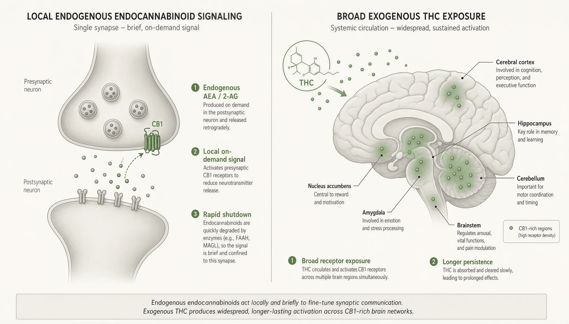
Spatial precision versus whole-brain exposure
Normal endocannabinoid signalling is local. Often very local. In one of the classic forms of ECS activity, a postsynaptic neuron becomes active, intracellular calcium rises, and that neuron synthesizes AEA or 2-AG on demand. The lipid messenger then travels backward across the synapse and activates presynaptic CB1 receptors, lowering the probability of neurotransmitter release. This retrograde process underlies depolarization-induced suppression of inhibition and excitation, worked out in late-1990s and early-2000s electrophysiology by researchers including Bradley Alger, Beat Lutz, Vincenzo Di Marzo, Tiziana Bisogno, Daniele Piomelli, and George Kunos, with important mechanistic clarification from Stella and Castillo as well. The point is not historical trivia. The point is scale: the endogenous signal is generated where needed, when needed, and usually at specific synapses.
THC does not respect that precision. Once inhaled or ingested and absorbed into the circulation, it reaches many CB1-rich regions at once: cortex, hippocampus, basal ganglia, cerebellum, amygdala, and others. CB1, cloned by Lisa Matsuda and colleagues in 1990, is one of the most abundant G protein-coupled receptors in the brain. That dense expression is why THC can alter memory, time perception, motor control, salience, appetite, and anxiety in one dose. Endocannabinoids can also influence those functions, but they usually do so by tuning ongoing circuits rather than bathing multiple networks at once in an external agonist.
This is where popular “deficient ECS” language goes wrong. AEA and 2-AG are not nutritional supplements the brain passively receives. They are event-driven signals. They appear in response to cellular activity and are shaped by local enzymatic machinery. 2-AG is usually the quantitatively dominant endocannabinoid in the brain and acts as a full agonist at CB1 in many systems; AEA is typically present at lower concentrations and is a partial agonist. Those are not interchangeable molecules. They differ in abundance, receptor efficacy, biosynthetic pathways, and timing.
THC flattens some of that selectivity. Instead of one active synapse transiently suppressing one presynaptic input, an exogenous cannabinoid can activate CB1 receptors across many active and inactive circuits alike. That is not “supporting homeostasis” in any simple sense. It is imposing a broad signal on a network designed for spatially constrained feedback.
CBD differs from THC here, but not in a way that rescues the simplistic story. CBD has low direct affinity for CB1 and CB2 at physiologically relevant concentrations and may act through several mechanisms, including negative allosteric modulation at CB1 in some models, TRPV1, 5-HT1A, adenosine-related pathways, ion channels, and nuclear receptors such as PPAR-gamma. So even when people describe CBD as “supporting the ECS,” that is often more slogan than mechanism. The pharmacology is real. The tidy explanation usually is not.
Signal duration and metabolism
Endocannabinoid signals are supposed to end fast. That is one of their defining features. After release, AEA is primarily hydrolyzed by fatty acid amide hydrolase, or FAAH. Brain 2-AG is mainly terminated by monoacylglycerol lipase, or MAGL; Nomura and colleagues reported in 2011 that MAGL accounts for about 85% of brain 2-AG hydrolysis activity in mice, with ABHD6 and ABHD12 contributing smaller shares. In other words, the ECS comes with built-in shutdown machinery.
THC is harder for the system to switch off. It is not synthesized on demand at the synapse, and it is not terminated by FAAH or MAGL in the way endogenous ligands are. Its pharmacokinetics depend on route of administration, dose, tissue distribution, and metabolism in the liver, not on the tight local off-switches that govern AEA and 2-AG. Because THC is lipophilic, it partitions into fatty tissues and can persist beyond the moment of subjective intoxication. The resulting receptor exposure is longer and less spatially disciplined than ordinary ECS signalling.
That difference in timing changes function. In physiological retrograde signalling, CB1 activation briefly suppresses neurotransmitter release, helping shape synaptic plasticity and circuit gain. With exogenous THC, CB1 activation can be stronger in some regions, longer-lasting, and detached from the original neural event that would have triggered endocannabinoid production. The result is not just more signalling. It is signalling with the wrong geometry and duration.
This is one reason enzyme inhibition is not equivalent to taking THC, and why “boosting your endocannabinoids” should not be treated as inherently benign. FAAH inhibitors once looked attractive because they seemed likely to amplify endogenous signalling only where and when endocannabinoids were already being produced. But even that approach turned out to be more complicated and risky than early rhetoric suggested. The BIA 10-2474 Phase I trial in France caused severe toxicity in 2016. That disaster likely reflected off-target effects rather than a clean demonstration that FAAH itself is dangerous to inhibit, but it shattered the lazy assumption that increasing endocannabinoid tone is automatically safe because it sounds more “natural.”
Rimonabant showed the other side of the same lesson. This CB1 inverse agonist produced meaningful weight loss in the RIO-Europe trial published by Van Gaal and colleagues in 2005, with 6.6 kg loss after one year in the 20 mg group versus 1.8 kg with placebo. It was also associated with psychiatric adverse effects and was withdrawn. The ECS is not a harmless balance dial. Push it too far in either direction and harm follows.
Tolerance, receptor downregulation, and adaptation
Repeated THC exposure changes the system. That is the central clinical point, and it is often softened or skipped in consumer-facing explanations.
CB1 receptors do not simply sit there and keep responding identically. With repeated agonist exposure, they can become desensitized, internalized, and downregulated. At the cellular level, receptor signalling weakens; at the systems level, users develop tolerance to at least some effects. This has been shown across animal studies, human imaging work, and postmortem receptor analyses. The pattern is not uniform. Tolerance develops more strongly for some effects than others, and receptor adaptation differs by brain region.
That regional unevenness matters. CB1 expression is especially high in cortex, hippocampus, basal ganglia, cerebellum, and limbic structures, but repeated THC does not produce identical adaptation in each area. Human PET studies and preclinical work suggest downregulation can be prominent in cortical regions and hippocampus, with somewhat different recovery trajectories across the brain after abstinence. That helps explain why tolerance may emerge to memory impairment, subjective intoxication, tachycardia, sleep effects, or appetite stimulation at different rates and to different degrees.
This is not the body calmly “adjusting to more support.” It is a receptor system compensating for overstimulation. If endogenous endocannabinoids are released briefly at selected synapses, the presynaptic neuron can recover once FAAH and MAGL clear the signal. But if THC repeatedly activates CB1 across broad regions, neurons adapt by making the receptor less available or less responsive. That adaptation is one reason heavy use can produce diminished acute effects over time and withdrawal symptoms when exposure stops. The system has been reset around the drug.
The same logic is why the phrase “supplements a deficient ECS” should be treated skeptically outside narrow research contexts. There are hypotheses about endocannabinoid deficiency in conditions such as migraine, fibromyalgia, and irritable bowel syndrome, but these remain hypotheses, not settled clinical doctrine. More importantly, even if some disorders involve altered endocannabinoid tone, inhaled or ingested cannabis is still not a precise replacement for local AEA or 2-AG signalling. It is a blunt intervention into a dynamic feedback network.
That does not mean cannabinoids have no therapeutic value. They do. Epidiolex, a purified CBD medicine, reduced convulsive seizure frequency by 43.9% versus 21.8% with placebo in Dravet syndrome in Devinsky et al. 2017, and reduced drop seizures in Lennox-Gastaut syndrome in Thiele et al. 2018. Nabiximols has evidence for multiple sclerosis spasticity in some jurisdictions. But successful cannabinoid therapeutics do not prove that cannabis simply restores natural ECS function. Usually the opposite lesson is more accurate: clinical benefit comes from carefully exploiting, and trying to control, a system that exogenous cannabinoids can just as easily perturb.
Clinical relevance: where ECS-targeted medicine has worked, and where it has failed
The jump from receptor pharmacology to actual treatment is where endocannabinoid science becomes more interesting and less forgiving. It is one thing to show that CB1 receptors regulate neurotransmitter release, that 2-AG is produced on demand, or that FAAH and MAGL terminate signalling. It is another to turn those facts into medicines that reliably help patients without causing damage somewhere else. The clinical record shows both outcomes. A few ECS-linked therapies have clear value. Others looked elegant on paper and collapsed in practice.
That distinction matters because “the ECS” is often used as a catch-all justification for broad cannabis claims. It should not be. The existence of an endogenous signalling network does not mean every cannabinoid product improves that network, and it does not mean that direct or indirect manipulation is automatically safe. If anything, the history of ECS-targeted medicine argues the opposite: this system is biologically powerful, widely distributed, and easy to perturb in ways that help one symptom while worsening another.
Approved or evidence-backed uses: epilepsy, antiemesis, pain, MS spasticity
The strongest modern example of a cannabinoid-related medicine succeeding is epilepsy, specifically purified cannabidiol. Epidiolex is not “cannabis” in the broad popular sense. It is a standardized pharmaceutical formulation of CBD, tested in randomized controlled trials and approved for seizures associated with Dravet syndrome, Lennox-Gastaut syndrome, and tuberous sclerosis complex. That is a real therapeutic success, but a narrow one.
The key trials were not subtle. In Dravet syndrome, Devinsky et al. published in The New England Journal of Medicine in 2017 that the cannabidiol group had a 43.9% reduction in convulsive-seizure frequency, compared with 21.8% in the placebo group. In Lennox-Gastaut syndrome, Thiele et al. reported in NEJM in 2018 that median drop-seizure frequency fell 41.9% with 20 mg/kg/day CBD and 37.2% with 10 mg/kg/day, versus 17.2% with placebo. Those are clinically meaningful effects in severe epilepsies that are often resistant to treatment.
This does not prove that CBD “supports the ECS” in some general wellness sense. In fact, CBD’s mechanism in epilepsy is still not fully pinned to classic CB1/CB2 signalling. CBD has low direct affinity for CB1 and CB2 at typical therapeutic concentrations. Its actions seem to involve a wider pharmacology that may include TRPV channels, GPR55, adenosine signalling, intracellular calcium regulation, and other targets. The lesson is simple: a cannabinoid can become a useful drug without acting as a clean, direct ECS agonist.
Antiemesis is another area where cannabinoid pharmacology has real clinical footing. The National Academies of Sciences, Engineering, and Medicine (NASEM) concluded in 2017 that there is conclusive or substantial evidence that cannabis or cannabinoids are effective for chemotherapy-induced nausea and vomiting in adults. That evidence base was built largely on older synthetic THC-related drugs such as dronabinol and nabilone rather than on modern dispensary-style products. Again, that distinction matters. The evidence is for particular agents, in a particular setting, with known dosing.
Mechanistically, this effect makes sense. Cannabinoid signalling influences emesis pathways in the brainstem and gut, even though CB1 expression in cardiorespiratory centers is relatively sparse compared with many other brain regions. But “makes sense” is not enough; antiemetic use earned support because controlled trials repeatedly showed benefit. That remains the standard.
Pain is more complicated. NASEM found substantial evidence that cannabis or cannabinoids are effective for chronic pain in adults, but that statement needs context. The evidence is heterogeneous, and “chronic pain” covers many different conditions with different mechanisms. Neuropathic pain has generally shown a stronger signal than nociceptive pain. Short-term symptom improvement is easier to demonstrate than durable functional improvement. Trials are often small, formulations vary, blinding is difficult because psychoactive effects can reveal treatment assignment, and adverse events are common.
So yes, there is evidence of analgesic benefit. No, that does not mean cannabinoids are universally effective pain medicines. It means there is a signal worth taking seriously, especially in selected chronic pain populations, while also acknowledging tradeoffs: dizziness, sedation, cognitive effects, psychiatric risks in susceptible patients, dependence risk with THC-rich products, and uncertain long-term outcomes. This is not an area for slogans.
Multiple sclerosis spasticity sits somewhere between success and partial success. Nabiximols, an oromucosal spray containing roughly equal amounts of THC and CBD, has evidence for improving patient-reported spasticity symptoms in MS and is approved in several countries for this indication. NASEM in 2017 judged that there is substantial evidence that oral cannabinoids improve patient-reported MS spasticity symptoms, though objective clinician-measured spasticity outcomes have been less consistently impressive.
That gap between patient-reported benefit and harder endpoint performance is not trivial. It may reflect real symptomatic relief that standard scales fail to capture, expectancy effects, psychoactive confounding, or some mix of all three. The fairest reading is that nabiximols and some oral cannabinoid preparations can help selected patients with refractory MS spasticity, but they are not a cure, not uniformly effective, and not proof that mixed cannabinoid products broadly “restore balance” across the nervous system.
The broader point across epilepsy, antiemesis, pain, and MS spasticity is this: evidence-backed cannabinoid medicine is specific, not general. Approved pharmaceuticals and supported indications exist. Grander claims usually outrun the data.
The rimonabant lesson: blocking CB1 can backfire
If one side of ECS medicine is benefit, the other is the reminder that this system is woven into mood, appetite, reward, stress response, and cognitive processing. Rimonabant made that painfully clear.
Rimonabant was a CB1 inverse agonist developed for obesity and metabolic disease. The rationale looked strong. CB1 signalling promotes appetite and participates in energy balance. Block it, and food intake should fall. Weight should drop. Metabolic markers might improve. And that is, in part, exactly what happened.
In the 2005 RIO-Europe trial led by Luc Van Gaal and colleagues, published in The Lancet, one-year weight loss was 6.6 kg in the rimonabant 20 mg group, compared with 1.8 kg with placebo. Waist circumference, lipid measures, and other cardiometabolic parameters also improved. On a narrow metabolic reading, the drug worked.
But CB1 is not confined to appetite circuits. It is one of the most abundant GPCRs in the brain, heavily represented in regions involved in emotion, reward, and stress responsivity. Blocking that signalling system systemically was never likely to be metabolically selective in real patients. Depression, anxiety, and suicidality emerged as serious adverse effects. The European Medicines Agency ultimately recommended suspension, and rimonabant was withdrawn.
This was not a minor setback. It was a conceptual warning. The ECS is often described as a homeostatic regulator, but that phrase can mislead people into thinking that less ECS signalling is bad in one domain and more is automatically good in another. Biology is not that tidy. Tonic and phasic cannabinoid signalling influence many circuits at once. A drug that pushes one node in the desired direction may destabilize another.
Rimonabant also exposed the danger of treating CB1 as if it were just an appetite switch. It is a network receptor in a network system. You can get weight loss by turning it down. You may also get psychiatric toxicity. Both findings are true, and medicine has to live with both.
FAAH inhibitors, BIA 10-2474, and the risks of elegant theories
After the rimonabant experience, indirect ECS modulation began to look more attractive than blunt receptor blockade or activation. Instead of pushing CB1 with THC-like agonists or suppressing it with antagonists, why not let the body’s own ligands do the work? That was the appeal of FAAH inhibition.
The theory was elegant. Anandamide is made on demand and rapidly broken down, largely by FAAH. Inhibit FAAH, raise anandamide levels, and perhaps enhance endogenous signalling only where and when the system is already active. That should, in principle, preserve some spatial and temporal specificity that exogenous cannabinoids lack. It promised analgesia and anxiolysis with fewer psychoactive effects than direct CB1 agonists.
For a while, this looked plausible. Several FAAH inhibitors entered development. Some early human studies did not reveal obvious catastrophic toxicity. The whole strategy seemed like a smarter, more physiological way to modulate the ECS.
Then came BIA 10-2474.
In 2016, a phase 1 trial in Rennes, France, testing the FAAH inhibitor BIA 10-2474, resulted in severe neurologic injury in multiple healthy volunteers and one death. The event shocked the field, and rightly so. A mechanism framed as subtle and endogenous had produced devastating toxicity at the first-in-human stage.
The exact reasons remain debated in detail, but the broad lessons are clear. First, “raising endocannabinoids” is not a synonym for safety. Second, drug effects cannot be inferred from target labels alone. BIA 10-2474 may have had problematic off-target actions, and its toxicity cannot simply be generalized to all FAAH inhibitors. That point matters because other FAAH inhibitors had not shown the same catastrophic pattern. Still, the disaster exposed a recurring mistake in translational pharmacology: elegant pathway logic can create false confidence.
The ECS invites that kind of overconfidence because its endogenous ligands are local, transient, and rapidly terminated. Intervening upstream or downstream can look gentler than it really is. Change degradation kinetics, and you may change signalling in tissues, compartments, or time windows that preclinical models did not capture. The same warning applies to MAGL inhibition. Since MAGL accounts for about 85% of brain 2-AG hydrolysis, according to Nomura et al. in Nature Chemical Biology in 2011, inhibiting it is not a minor tweak. It is a major intervention in a dominant lipid signalling pathway.
The BIA 10-2474 case did not prove that all enzyme-targeted ECS drugs are doomed. It proved something more important: the endocannabinoid system is a real pharmacologic control layer, and interfering with it can produce large effects, good or bad. That is exactly why the field deserves both scientific seriousness and clinical restraint.
- Clear successes
- Purified CBD in severe epilepsies; antiemetic use; some MS spasticity benefit
- Supported but variable
- Chronic pain in selected adults
- Major failures
- Rimonabant psychiatric toxicity; BIA 10-2474 severe toxicity
- Overall lesson
- The ECS is medically useful to target, but easy to perturb in harmful ways
The current state of ECS-targeted medicine is therefore mixed but not confusing. Some interventions have worked and earned their place. Epidiolex is one. Nabiximols, in certain MS settings, is another qualified example. Cannabinoids for chemotherapy-related nausea and selected chronic pain conditions have evidentiary support, though the details matter more than the headlines. At the same time, rimonabant showed that blocking CB1 can damage mental health, and BIA 10-2474 showed that indirect enhancement of endocannabinoid tone is not automatically benign.
That is the mature view of the field. The ECS is medically important because it can be manipulated. It is medically dangerous for exactly the same reason.
The evidence gaps and disputed claims around the ECS
The endocannabinoid system is real biology, not wellness poetry. CB1 was cloned by Lisa Matsuda and colleagues in Nature in 1990. CB2 followed with Munro and colleagues in 1993. Anandamide was identified by Devane, Hanuš, Breuer, Mechoulam and coauthors in 1992, and 2-AG was established as an endocannabinoid in 1995 by Mechoulam’s and Sugiura’s groups. Since then, the field has mapped a genuine lipid signalling network: endocannabinoids are synthesized on demand from membrane precursors, often travel backward across the synapse, and are rapidly shut down by enzymes such as FAAH and MAGL. That is a much more precise picture than the usual “THC binds CB1” simplification.
Still, a real system can be surrounded by weak claims. That has happened here. The ECS is often drafted into explanations that outrun the data, especially in consumer-facing CBD and cannabis content. Editorially, three points are defensible. First, “clinical endocannabinoid deficiency” is an interesting hypothesis, not an established diagnosis. Second, the entourage effect is pharmacologically plausible, but the human evidence is thin relative to how confidently it is discussed. Third, animal studies remain indispensable for mechanism and very shaky grounds for specific, consumer-level promises about how a cannabinoid product will feel or perform in people.
Clinical endocannabinoid deficiency: hypothesis versus proof
Clinical endocannabinoid deficiency is a research hypothesis, not an established diagnosis. Preliminary evidence
Clinical endocannabinoid deficiency (CECD) A proposed framework suggesting that some disorders may involve low endocannabinoid tone. The article treats it as an interesting but unproven hypothesis.
The clinical endocannabinoid deficiency, or CECD, hypothesis is most closely associated with Ethan Russo, who argued in the early 2000s that disorders such as migraine, fibromyalgia, and irritable bowel syndrome might reflect low endocannabinoid tone. The idea has intuitive appeal because those conditions can involve pain sensitivity, stress reactivity, altered gut function, and other processes in which the ECS participates. It also fits the broader observation that the ECS helps adjust set points rather than acting as a simple on-off switch.
Interesting is not the same as established.
The main problem is proof. There is no validated clinical test that can diagnose CECD in routine practice. Anandamide and 2-AG fluctuate by tissue, time, diet, stress state, inflammation, menstrual phase, and sampling method. Peripheral blood values do not straightforwardly tell you what is happening at a synapse in the hippocampus, amygdala, dorsal horn, or enteric nervous system. Even when studies report altered endocannabinoid levels in a disease state, causation is unresolved. Low tone may contribute to illness, reflect illness, or represent compensation for some other disturbance.
The receptor side is no easier. CB1 and CB2 expression can shift with chronic stress, injury, obesity, inflammation, drug exposure, and disease. CB2 in the central nervous system is a good example of why simplistic maps fail: it is mainly associated with immune cells and tissues, but low-level neural expression in some conditions remains context-dependent and debated. So even if “deficiency” were the right frame in one tissue, it might be wrong in another.
There is also a conceptual trap here. The ECS does not always restore health when pushed upward. Rimonabant, a CB1 inverse agonist, produced meaningful weight loss in the 2005 RIO-Europe trial by Van Gaal et al. — 6.6 kg at one year with 20 mg versus 1.8 kg with placebo — but was withdrawn because psychiatric adverse effects were serious. The lesson is not just that blocking CB1 can be harmful. It is that the ECS is deeply woven into mood, motivation, feeding, and stress circuits. Intervening in it can help, harm, or do both at once. Likewise, FAAH inhibition looked elegant on paper because it seemed likely to raise anandamide where it was needed, yet the BIA 10-2474 phase 1 trial in France caused severe toxicity. “Boosting endocannabinoids” is not a synonym for safety.
So the right position is restrained: CECD is a useful research hypothesis and a provocative way to organize some observations, but it is not a proven diagnostic framework and should not be presented as settled clinical fact.
The entourage effect: plausible, popular, under-tested
The entourage effect is one of the most over-claimed ideas in cannabinoid medicine. In its strongest public form, it says mixtures of cannabinoids and terpenes consistently work better than isolated compounds because the plant’s chemistry produces cooperative effects. The first half of that statement is plausible. The second half is often asserted far beyond the evidence.
Pharmacologically, interactions are easy to imagine. THC is a partial agonist at CB1 and CB2. CBD has low direct affinity for those receptors at typical concentrations and appears to act through a messier profile that may include negative allosteric effects at CB1, TRPV1, 5-HT1A, adenosine-related signalling, PPAR-gamma, and ion channels. CBG has weak CB1/CB2 activity and other targets, including alpha-2 adrenergic and TRP-family interactions. On top of that, terpenes may have their own receptor and membrane effects. So yes, multi-compound interaction is pharmacologically credible.
But credibility is not confirmation.
Human evidence for broad entourage claims is limited, heterogeneous, and often confounded by dosing, route of administration, expectation effects, and product variability. Some whole-plant or extract-based medicines have evidence in specific conditions; nabiximols has data for multiple-sclerosis spasticity in some jurisdictions. That does not validate every “full-spectrum works better” claim. Nor does the success of purified CBD in epilepsy argue against isolates as a class. In NEJM, Devinsky et al. 2017 showed a 43.9% reduction in convulsive-seizure frequency with cannabidiol in Dravet syndrome versus 21.8% with placebo. Thiele et al. 2018 found median drop-seizure reductions of 41.9% with 20 mg/kg/day CBD and 17.2% with placebo in Lennox-Gastaut syndrome. Those are purified-drug data, not proof that complex extracts are superior.
My position is simple: the entourage effect should be treated as a family of testable interaction hypotheses, not a default truth. Some combinations may be additive, some antagonistic, some irrelevant, and some dependent on dose and indication. The phrase is useful only if it points toward actual pharmacology. Too often it functions as a rhetorical shortcut.
What animal models can and cannot tell us
Without animal work, the ECS would still be largely invisible. Retrograde signalling through endocannabinoids was clarified through electrophysiology in the late 1990s and early 2000s by researchers including Bradley Alger, Daniele Piomelli’s collaborators, and Alfonso Castillo’s group. Knockout mice, receptor autoradiography, microdialysis, and enzyme-inhibition studies established many basics of receptor distribution and ligand turnover. Nomura et al. showed in 2011 that MAGL accounts for about 85% of brain 2-AG hydrolysis activity in mouse brain. Those mechanistic gains matter.
They are not the same thing as reliable product predictions in humans.
Rodent studies can show that a cannabinoid reduces anxiety-like behavior in one paradigm, increases it in another, suppresses inflammatory pain, alters fear extinction, changes feeding, modifies seizure threshold, or shifts social behavior. The translation problem is obvious: “anxiety-like behavior” in elevated plus maze time is not generalized anxiety disorder. A chemically induced colitis model is not the full lived reality of inflammatory bowel disease. Mouse strain, sex, housing stress, timing, route of administration, and dose can all change the result. Cannabinoids also show biphasic effects. A low dose can do one thing; a higher dose can reverse it.
That is why animal evidence is strongest when used for mechanism: identifying receptor involvement, mapping circuits, separating AEA from 2-AG functions, or testing how FAAH versus MAGL inhibition changes signalling. It is much weaker when used to support direct consumer-facing statements such as “CBD calms the nervous system,” “CBG sharpens focus,” or “a terpene blend enhances THC in a predictable way.” Those claims usually skip several levels of uncertainty.
A sober reading of the ECS literature does not diminish the field. It improves it. The system is important, clinically relevant, and biologically rich. But it is not a blank check for speculation.
Why the ECS matters for every cannabinoid article on this wiki
The endocannabinoid system is the reference frame for almost every serious claim made about cannabinoids. Without it, THC gets reduced to “the psychoactive one,” CBD to “the calming one,” and CBG to “the minor one with promise,” which is exactly how bad cannabis writing starts. The ECS is not a cannabis-processing module that evolved for the plant. It is an endogenous lipid signalling network discovered partly because cannabis researchers followed the pharmacology. CB1 was cloned by Lisa Matsuda and colleagues in Nature in 1990; CB2 followed with Munro et al. in 1993; anandamide was identified by Devane, Hanuš, and colleagues in 1992; 2-AG was established as an endocannabinoid in 1995 by Mechoulam’s and Sugiura’s groups. That timeline matters because it shows the direction of causality: cannabis helped reveal the system, but the system is native to the body.
That distinction changes how this wiki reads every cannabinoid article linked from here. Endocannabinoids are made on demand from membrane lipids, act locally, often travel backward across synapses from postsynaptic to presynaptic cells, and are shut down fast by enzymes such as FAAH and MAGL. THC does not reproduce that pattern neatly. It can mimic parts of it, but with different timing, broader tissue exposure, different efficacy, and often longer persistence. So when a product, strain, or isolated compound is said to “support the ECS,” the first question should be: by what mechanism, in what tissue, at what dose, by which route, and with what evidence in humans?
Reading THC, CBD, and CBG through ECS biology
ECS biology gives readers a way to sort compounds by mechanism instead of marketing categories. THC makes the easiest starting point because it is a partial agonist at CB1 and CB2, and CB1 is highly expressed in cortex, hippocampus, basal ganglia, cerebellum, and limbic regions. That receptor geography helps explain intoxication, memory disruption, altered time perception, appetite effects, and motor changes. It also helps explain what THC does not usually do: because CB1 expression is sparse in brainstem cardiorespiratory centers, cannabis does not produce the classic fatal respiratory depression pattern seen with opioid overdose.
CBD is harder, and that is exactly why ECS literacy matters. CBD has low direct affinity for CB1 and CB2 at physiologically relevant concentrations, so simplistic “CBD activates the ECS” language is usually wrong. Its actions appear to involve a messier set of targets and modulators, including possible negative allosteric modulation at CB1, TRPV1, 5-HT1A, adenosine-related signalling, PPAR-gamma, and ion-channel effects. Some contexts suggest FAAH-related effects, but that does not make CBD a clean endocannabinoid booster. Readers should keep one fact in view: the evidence base for purified prescription CBD is not the same thing as the evidence base for retail CBD products. In NEJM, Devinsky et al. 2017 reported a 43.9% reduction in convulsive-seizure frequency with cannabidiol in Dravet syndrome versus 21.8% with placebo; Thiele et al. 2018 found a 41.9% drop in Lennox-Gastaut drop seizures at 20 mg/kg/day versus 17.2% with placebo. Those are real clinical signals. They do not validate every broad CBD claim attached to sleep, mood, inflammation, or “balance.”
CBG should be read even more cautiously. It has weak or low-affinity interactions at CB1 and CB2 and also engages non-cannabinoid targets such as alpha-2 adrenergic receptors and TRP channels. That makes pharmacological action plausible. It does not make effect claims mature. Human clinical data remain thin.
The same logic extends beyond cannabinoids to terpenes, formulations, and routes of administration. A terpene claim only matters if the compound reaches relevant concentrations in vivo. An edible, inhaled dose, and oral tincture can produce very different exposure curves and metabolites. 11-hydroxy-THC after oral dosing is the classic example. ECS knowledge turns “this feels different” into a pharmacokinetic question rather than a mystical one.
What receptor affinity does not tell you
Affinity charts are useful, but they are not outcome charts. A binding number says something about how strongly a compound interacts with a target under specific assay conditions. It does not tell you whether the compound is an agonist, partial agonist, antagonist, inverse agonist, allosteric modulator, or functionally irrelevant at real human exposure levels. It also does not tell you where in the body the target is expressed, whether the compound gets there, how fast it arrives, how long it persists, what metabolites form, or what competing ligands are present.
That is especially important in the ECS because the endogenous system is kinetic and local. Anandamide and 2-AG are not interchangeable “natural cannabinoids.” AEA is generally lower in tissue abundance and acts as a partial agonist at CB1. 2-AG is usually the dominant brain endocannabinoid and acts as a full agonist at CB1 and CB2 in many systems. Their shutdown is different too: FAAH primarily degrades AEA, while MAGL accounts for about 85% of brain 2-AG hydrolysis, according to Nomura et al. 2011. If a compound changes one arm of that network but not the other, the physiological result may be quite specific.
Clinical history makes the point even sharper. Rimonabant, a CB1 inverse agonist, produced weight loss in the 2005 RIO-Europe trial—6.6 kg after one year at 20 mg versus 1.8 kg with placebo—yet it was withdrawn because psychiatric adverse effects were serious. So “targeting CB1 works” was both true and dangerously incomplete. The failed FAAH inhibitor BIA 10-2474 was another warning: increasing endocannabinoid tone is not automatically gentle or safe.
How to interpret product and strain claims more skeptically
This wiki uses the ECS as a filter against inflated claims. If a label says a product is “for focus,” “for inflammation,” or “for sleep,” readers should ask whether the claim is tied to controlled human evidence, a plausible mechanism, or just a familiar narrative wrapped around a chemovar name. Strain names are especially poor proxies for pharmacology. Chemotype, dose, route, user tolerance, metabolism, and setting usually matter more.
The same skepticism applies to entourage-effect rhetoric. Multi-compound interactions are pharmacologically plausible. That much is fair. But direct human evidence is far weaker than the confidence of the marketing language built around it. A terpene profile does not guarantee a predictable subjective or therapeutic outcome. Nor does receptor talk rescue a weak claim. Context matters: lab certificate quality, cannabinoid ratios, contaminant testing, oral bioavailability, inhalation topography, first-pass metabolism, and individual variation in enzymes and prior exposure all shape effects.
This is the hub logic of the wiki. ECS biology lets readers move from slogans to mechanisms, from isolated binding data to whole-system interpretation, and from cannabinoid myths to evidence-weighted reading. Every cannabinoid article that follows—THC, CBD, CBG, CBN, CBC, delta-8-THC, THCV, and others—makes more sense once you see the same underlying rule: cannabis compounds do not simply “activate” a dormant system. They perturb an active one, sometimes subtly, sometimes forcefully, and sometimes in ways the body did not design for.
Legal, medical, and practical cautions when discussing the ECS
The endocannabinoid system is a real signalling network, not a wellness metaphor and not a blank check for medical claims. That distinction matters because ECS language is often used to jump from receptor biology to sweeping statements about what cannabis, CBD, or other cannabinoids “should” do in the body. The science does not support that shortcut. Discussion here is informational only and should not be read as personal medical advice, diagnosis, or a recommendation to use any ECS-targeting compound.
Medical-claim boundaries
Explaining mechanism is fair. Claiming disease treatment requires clinical evidence. Those are different standards.
For example, it is accurate to say that CB1 was cloned by Matsuda et al. in 1990, CB2 by Munro et al. in 1993, anandamide was identified by Devane et al. in 1992, and 2-AG was established as an endocannabinoid in 1995 by Mechoulam and colleagues and by Sugiura and colleagues. It is also accurate to say that endocannabinoids are produced on demand, often act retrogradely, and are rapidly broken down by FAAH and MAGL, with MAGL responsible for about 85% of brain 2-AG hydrolysis in mouse brain in Nomura et al. 2011. None of that, by itself, proves that a given cannabinoid product treats anxiety, pain, insomnia, inflammation, or any other condition.
The medical record is mixed, not uniformly positive. There are genuine successes. Purified prescription cannabidiol has regulatory approval for seizures associated with Lennox-Gastaut syndrome, Dravet syndrome, and tuberous sclerosis complex. In Devinsky et al. 2017, convulsive-seizure frequency in Dravet syndrome fell 43.9% with cannabidiol versus 21.8% with placebo. In Thiele et al. 2018, drop seizures in Lennox-Gastaut syndrome fell 41.9% with 20 mg/kg/day CBD versus 17.2% with placebo. Those are meaningful data. They do not validate retail CBD products, and they do not mean CBD is broadly proven for every ECS-linked disorder.
There are failures and harms too. Rimonabant, a CB1 inverse agonist, produced weight loss in RIO-Europe in 2005 but was withdrawn because psychiatric adverse effects were serious. The BIA 10-2474 FAAH inhibitor trial in France caused severe toxicity. That is the practical lesson many popular explainers miss: manipulating the ECS can help, do nothing, or cause harm. “Natural” and “endocannabinoid-boosting” are not safety guarantees.
Jurisdictional variation in cannabis law
Cannabis law is fragmented and changes often. A product may be legal in one country, illegal in the next, and heavily restricted in a third. Even within a single federal system, state, provincial, or territorial rules may diverge from national policy. That affects possession, prescribing, product standards, THC limits, driving laws, workplace testing, and what counts as lawful medical access.
This matters because ECS discussions often slide into practical use questions. Readers should assume nothing. Check current law in the relevant jurisdiction through official government sources, not old summaries, social media posts, or packaging claims. The scale of use does not settle legality either: UNODC estimated 228 million past-year cannabis users worldwide in 2022, and SAMHSA estimated 61.9 million past-year U.S. users aged 12 and older in 2022. Widespread use is a public-health fact. It is not legal advice.
Why mechanism does not equal treatment recommendation
Mechanistic plausibility is where many ECS discussions go off the rails. A receptor in a tissue does not mean a cannabinoid will improve a disease in that tissue. A pathway involved in homeostasis does not mean pushing it will restore health. The ECS regulates synaptic signalling, appetite, pain, immune tone, gastrointestinal motility, stress responses, and more. That breadth makes it scientifically interesting and clinically difficult.
THC does not simply “activate the ECS.” It perturbs it. Endocannabinoids are made locally, on demand, then cleared quickly by enzymes such as FAAH and MAGL. THC arrives from outside the system, reaches tissues on a different timescale, and persists longer. CBD is even less straightforward: low direct affinity at CB1 and CB2, possible negative allosteric modulation at CB1, TRPV1 and 5-HT1A actions, adenosine and ion-channel effects, and context-dependent FAAH-related findings. CBG is pharmacologically interesting, but clinically thin. Claims about an “entourage effect” remain plausible at the pharmacology level and underproven in direct human evidence.
So the safe intellectual rule is simple: mechanism can justify research. It cannot, on its own, justify treatment advice. For personal medical decisions, readers should rely on a qualified clinician and high-quality human data, not receptor diagrams.
References
- [1] Structure of a cannabinoid receptor and functional expression of the cloned cDNA. Nature, 1990. https://doi.org/10.1038/346561a0
- [2] Molecular characterization of a peripheral receptor for cannabinoids. Nature, 1993. https://doi.org/10.1038/365061a0
- [3] Isolation and structure of a brain constituent that binds to the cannabinoid receptor. Science, 1992. https://doi.org/10.1126/science.1470919
- [4] Monoacylglycerol lipase regulates a fatty acid network that promotes cancer pathogenesis. Nature Chemical Biology, 2011. https://doi.org/10.1038/nchembio.593
- [5] Effects of the cannabinoid-1 receptor blocker rimonabant on weight reduction and cardiovascular risk factors in overweight patients: 1-year experience from the RIO-Europe study. The Lancet, 2005. https://doi.org/10.1016/S0140-6736(05)67214-2
- [6] Trial of Cannabidiol for Drug-Resistant Seizures in the Dravet Syndrome. The New England Journal of Medicine, 2017. https://doi.org/10.1056/NEJMoa1611618
- [7] Cannabidiol in Patients with Seizures Associated with Lennox-Gastaut Syndrome. The New England Journal of Medicine, 2018. https://doi.org/10.1056/NEJMoa1714631
- [8] Cannabidiol (CBD) Critical Review Report. World Health Organization report, 2018. https://www.who.int/publications/m/item/cannabidiol-(cbd)-critical-review-report